5IQS

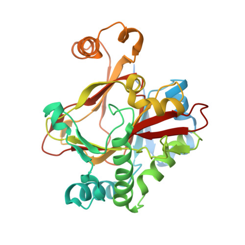
WelO5 bound to Fe(II), Cl, and 2-oxoglutarate


Experimental Data Snapshot

  • Method: X-RAY DIFFRACTION
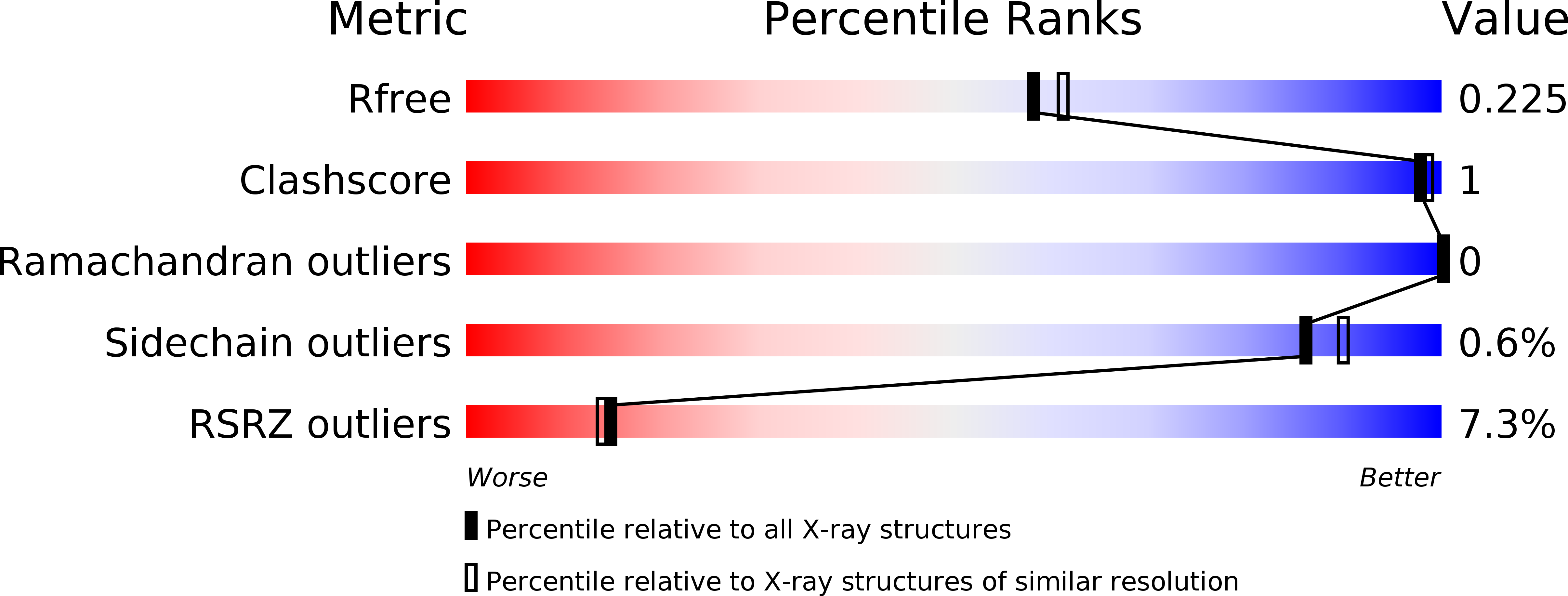
  • Resolution: 2.00 Å
  • R-Value Free: 0.219 
  • R-Value Work: 0.189 
  • R-Value Observed: 0.191 

wwPDB Validation   3D Report Full Report


This is version 1.4 of the entry. See complete history


Literature

Structural basis for halogenation by iron- and 2-oxo-glutarate-dependent enzyme WelO5.

Mitchell, A.J.Zhu, Q.Maggiolo, A.O.Ananth, N.R.Hillwig, M.L.Liu, X.Boal, A.K.

(2016) Nat Chem Biol 12: 636-640

  • DOI: https://doi.org/10.1038/nchembio.2112
  • Primary Citation of Related Structures:  
    5IQS, 5IQT, 5IQU, 5IQV

  • PubMed Abstract: 

    A 2.4-Å-resolution X-ray crystal structure of the carrier-protein-independent halogenase WelO5 in complex with its welwitindolinone precursor substrate, 12-epi-fischerindole U, reveals that the C13 chlorination target is proximal to the anticipated site of the oxo group in a presumptive cis-halo-oxo-iron(IV) (haloferryl) intermediate. Prior study of related halogenases forecasts substrate hydroxylation in this active-site configuration, but X-ray crystallographic verification of C13 halogenation in single crystals mandates that ligand dynamics must reposition the oxygen ligand to enable the observed outcome. S189A WelO5 produces a mixture of halogenation and hydroxylation products, showing that an outer-sphere hydrogen-bonding group orchestrates ligand movements to achieve a configuration that promotes halogen transfer.


  • Organizational Affiliation

    Department of Biochemistry and Molecular Biology, The Pennsylvania State University, University Park, Pennsylvania, USA.


Macromolecules
Find similar proteins by:  (by identity cutoff)  |  3D Structure
Entity ID: 1
MoleculeChains Sequence LengthOrganismDetailsImage
WelO5
A, B, C
315Hapalosiphon welwitschii UTEX B 1830Mutation(s): 0 
UniProt
Find proteins for A0A067YX61 (Hapalosiphon welwitschii UTEX B 1830)
Explore A0A067YX61 
Go to UniProtKB:  A0A067YX61
Entity Groups  
Sequence Clusters30% Identity50% Identity70% Identity90% Identity95% Identity100% Identity
UniProt GroupA0A067YX61
Sequence Annotations
Expand
  • Reference Sequence
Small Molecules
Ligands 3 Unique
IDChains Name / Formula / InChI Key2D Diagram3D Interactions
AKG
Query on AKG

Download Ideal Coordinates CCD File 
E [auth A],
H [auth B],
K [auth C]
2-OXOGLUTARIC ACID
C5 H6 O5
KPGXRSRHYNQIFN-UHFFFAOYSA-N
FE2
Query on FE2

Download Ideal Coordinates CCD File 
D [auth A],
G [auth B],
J [auth C]
FE (II) ION
Fe
CWYNVVGOOAEACU-UHFFFAOYSA-N
CL
Query on CL

Download Ideal Coordinates CCD File 
F [auth A],
I [auth B],
L [auth C]
CHLORIDE ION
Cl
VEXZGXHMUGYJMC-UHFFFAOYSA-M
Experimental Data & Validation

Experimental Data

  • Method: X-RAY DIFFRACTION
  • Resolution: 2.00 Å
  • R-Value Free: 0.219 
  • R-Value Work: 0.189 
  • R-Value Observed: 0.191 
  • Space Group: P 21 21 21
Unit Cell:
Length ( Å )Angle ( ˚ )
a = 70.757α = 90
b = 86.339β = 90
c = 141.312γ = 90
Software Package:
Software NamePurpose
HKL-2000data scaling
REFMACrefinement
PDB_EXTRACTdata extraction
autoSHARPphasing
HKL-2000data reduction

Structure Validation

View Full Validation Report


Entry History & Funding Information

Deposition Data


Funding OrganizationLocationGrant Number
National Institutes of Health/National Institute of General Medical Sciences (NIH/NIGMS)United StatesGM100011

Revision History  (Full details and data files)

  • Version 1.0: 2016-06-29
    Type: Initial release
  • Version 1.1: 2016-07-13
    Changes: Database references
  • Version 1.2: 2016-07-27
    Changes: Database references
  • Version 1.3: 2017-09-13
    Changes: Author supporting evidence, Derived calculations
  • Version 1.4: 2022-04-13
    Changes: Author supporting evidence, Database references, Derived calculations