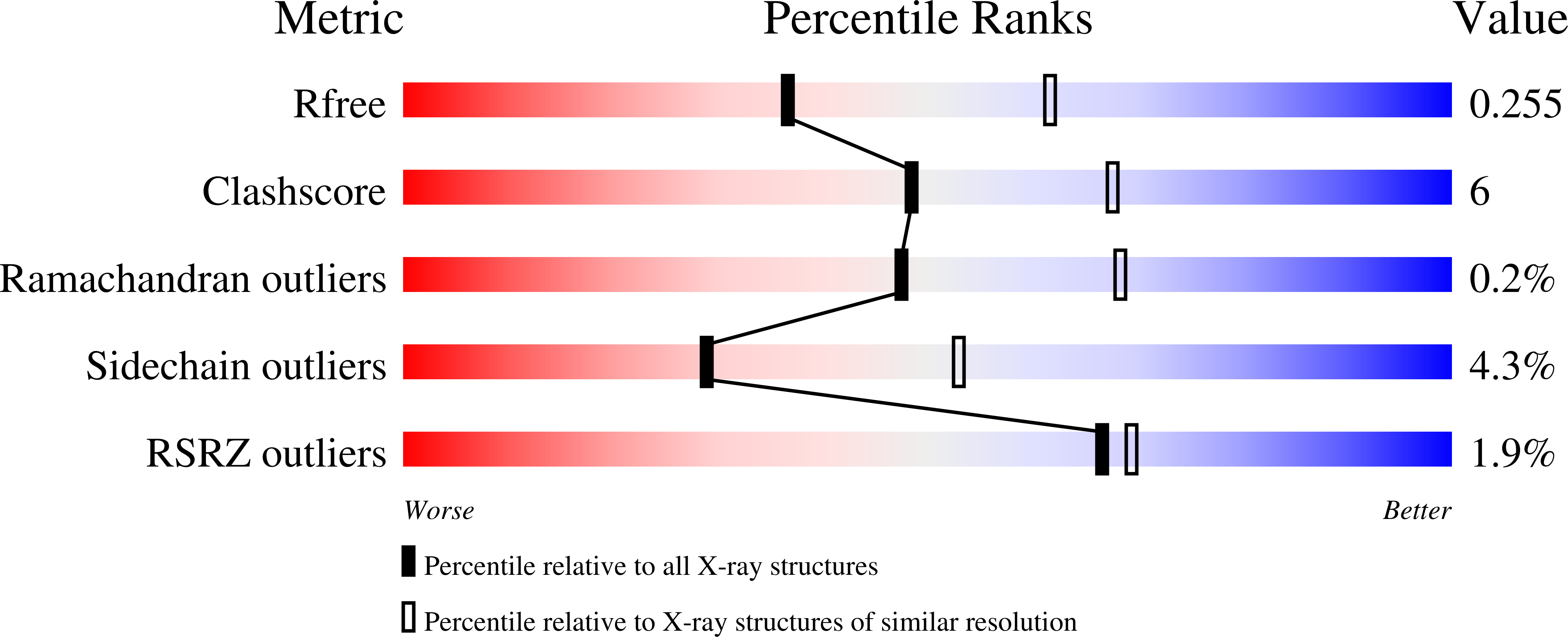
Crystal structure of truncated aspartate transcarbamoylase from Plasmodium falciparum.
Lunev, S., Bosch, S.S., Batista, F.d.A., Wrenger, C., Groves, M.R.(2016) Acta Crystallogr F Struct Biol Commun 72: 523-533
- PubMed: 27380369
- DOI: https://doi.org/10.1107/S2053230X16008475
- Primary Citation of Related Structures:
5ILQ - PubMed Abstract:
The de novo pyrimidine-biosynthesis pathway of Plasmodium falciparum is a promising target for antimalarial drug discovery. The parasite requires a supply of purines and pyrimidines for growth and proliferation and is unable to take up pyrimidines from the host. Direct (or indirect) inhibition of de novo pyrimidine biosynthesis via dihydroorotate dehydrogenase (PfDHODH), the fourth enzyme of the pathway, has already been shown to be lethal to the parasite. In the second step of the plasmodial pyrimidine-synthesis pathway, aspartate and carbamoyl phosphate are condensed to N-carbamoyl-L-aspartate and inorganic phosphate by aspartate transcarbamoylase (PfATC). In this paper, the 2.5 Å resolution crystal structure of PfATC is reported. The space group of the PfATC crystals was determined to be monoclinic P21, with unit-cell parameters a = 87.0, b = 103.8, c = 87.1 Å, α = 90.0, β = 117.7, γ = 90.0°. The presented PfATC model shares a high degree of homology with the catalytic domain of Escherichia coli ATC. There is as yet no evidence of the existence of a regulatory domain in PfATC. Similarly to E. coli ATC, PfATC was modelled as a homotrimer in which each of the three active sites is formed at the oligomeric interface. Each active site comprises residues from two adjacent subunits in the trimer with a high degree of evolutional conservation. Here, the activity loss owing to mutagenesis of the key active-site residues is also described.
Organizational Affiliation:
Department of Drug Design, Groningen Research Institute of Pharmacy, University of Groningen, Antonius Deusinglaan 1, 9700 AD Groningen, The Netherlands.


