


Crystal Structure of Human Jmjd2A in Complex with D-Threo-Isocitrate
Nowak, R., Kopec, J., Johansson, C., Szykowska, A., von Delft, F., Arrowsmith, C.H., Bountra, C., Edwards, A., Oppermann, U.To be published.
Experimental Data Snapshot
Entity ID: 1 | |||||
|---|---|---|---|---|---|
| Molecule | Chains | Sequence Length | Organism | Details | Image |
| LYSINE-SPECIFIC DEMETHYLASE 4A | 381 | Homo sapiens | Mutation(s): 0 EC: 1.14.11 | 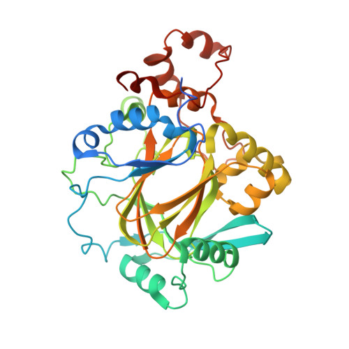 | |
UniProt & NIH Common Fund Data Resources | |||||
Find proteins for O75164 (Homo sapiens) Explore O75164 Go to UniProtKB: O75164 | |||||
PHAROS: O75164 GTEx: ENSG00000066135 | |||||
Entity Groups | |||||
| Sequence Clusters | 30% Identity50% Identity70% Identity90% Identity95% Identity100% Identity | ||||
| UniProt Group | O75164 | ||||
Sequence AnnotationsExpand | |||||
| |||||
| Ligands 5 Unique | |||||
|---|---|---|---|---|---|
| ID | Chains | Name / Formula / InChI Key | 2D Diagram | 3D Interactions | |
| N81 Query on N81 | E [auth A], L [auth B] | 3-carboxy-2,3-dideoxy-D-erythro-pentaric acid C6 H8 O7 ODBLHEXUDAPZAU-OKKQSCSOSA-N |  | ||
| DMS Query on DMS | F [auth A], M [auth B] | DIMETHYL SULFOXIDE C2 H6 O S IAZDPXIOMUYVGZ-UHFFFAOYSA-N |  | ||
| ZN Query on ZN | D [auth A], K [auth B] | ZINC ION Zn PTFCDOFLOPIGGS-UHFFFAOYSA-N |  | ||
| EDO Query on EDO | G [auth A] H [auth A] I [auth A] N [auth B] O [auth B] | 1,2-ETHANEDIOL C2 H6 O2 LYCAIKOWRPUZTN-UHFFFAOYSA-N |  | ||
| NI Query on NI | C [auth A], J [auth B] | NICKEL (II) ION Ni VEQPNABPJHWNSG-UHFFFAOYSA-N |  | ||
| Length ( Å ) | Angle ( ˚ ) |
|---|---|
| a = 100.93 | α = 90 |
| b = 148.91 | β = 90 |
| c = 57.05 | γ = 90 |
| Software Name | Purpose |
|---|---|
| PHENIX | refinement |
| XDS | data reduction |
| SCALA | data scaling |
| DIMPLE | phasing |
RCSB PDB (citation) is hosted by
RCSB PDB is a member of the

