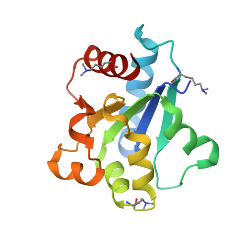
5FOR

Cryptic TIR


Experimental Data Snapshot

  • Method: X-RAY DIFFRACTION
  • Resolution: 2.50 Å
  • R-Value Free: 0.244 
  • R-Value Work: 0.188 
  • R-Value Observed: 0.191 

wwPDB Validation   3D Report Full Report


Ligand Structure Quality Assessment 


This is version 1.2 of the entry. See complete history


Literature

Structure of the Toll/Interleukin-1 Receptor (TIR) Domain of the B-cell Adaptor That Links Phosphoinositide Metabolism with the Negative Regulation of the Toll-like Receptor (TLR) Signalosome.

Halabi, S.Sekine, E.Verstak, B.Gay, N.J.Moncrieffe, M.C.

(2017) J Biol Chem 292: 652-660

  • DOI: https://doi.org/10.1074/jbc.M116.761528
  • Primary Citation of Related Structures:  
    5FOR

  • PubMed Abstract: 

    Ligand binding to Toll-like receptors (TLRs) results in dimerization of their cytosolic Toll/interleukin-1 receptor (TIR) domains and recruitment of post-receptor signal transducers into a complex signalosome. TLR activation leads to the production of transcription factors and pro-inflammatory molecules and the activation of phosphoinositide 3-kinases (PI3K) in a process that requires the multimodular B-cell adaptor for phosphoinositide 3-kinase (BCAP). BCAP has a sequence previously proposed as a "cryptic" TIR domain. Here, we present the structure of the N-terminal region of human BCAP and show that it possesses a canonical TIR fold. Dimeric BCAP associates with the TIR domains of TLR2/4 and MAL/TIRAP, suggesting that it is recruited to the TLR signalosome by multitypic TIR-TIR interactions. BCAP also interacts with the p85 subunit of PI3K and phospholipase Cγ, enzymes that deplete plasma membrane phosphatidylinositol 4,5-bisphosphate (PIP2), and these interactions provide a molecular explanation for BCAP-mediated down-regulation of inflammatory signaling.


  • Organizational Affiliation

    From the Department of Biochemistry, Cambridge University, Cambridge CB2 1GA, United Kingdom.


Macromolecules
Find similar proteins by:  (by identity cutoff)  |  3D Structure
Entity ID: 1
MoleculeChains Sequence LengthOrganismDetailsImage
PHOSPHOINOSITIDE 3-KINASE ADAPTER PROTEIN 1139Homo sapiensMutation(s): 0 
UniProt & NIH Common Fund Data Resources
Find proteins for Q6ZUJ8 (Homo sapiens)
Explore Q6ZUJ8 
Go to UniProtKB:  Q6ZUJ8
PHAROS:  Q6ZUJ8
GTEx:  ENSG00000155629 
Entity Groups  
Sequence Clusters30% Identity50% Identity70% Identity90% Identity95% Identity100% Identity
UniProt GroupQ6ZUJ8
Sequence Annotations
Expand
  • Reference Sequence
Small Molecules
Ligands 2 Unique
IDChains Name / Formula / InChI Key2D Diagram3D Interactions
I3C
Query on I3C

Download Ideal Coordinates CCD File 
C [auth A]5-amino-2,4,6-triiodobenzene-1,3-dicarboxylic acid
C8 H4 I3 N O4
JEZJSNULLBSYHV-UHFFFAOYSA-N
1PE
Query on 1PE

Download Ideal Coordinates CCD File 
B [auth A]PENTAETHYLENE GLYCOL
C10 H22 O6
JLFNLZLINWHATN-UHFFFAOYSA-N
Modified Residues  1 Unique
IDChains TypeFormula2D DiagramParent
MLY
Query on MLY
A
L-PEPTIDE LINKINGC8 H18 N2 O2LYS
Experimental Data & Validation

Experimental Data

  • Method: X-RAY DIFFRACTION
  • Resolution: 2.50 Å
  • R-Value Free: 0.244 
  • R-Value Work: 0.188 
  • R-Value Observed: 0.191 
  • Space Group: P 62 2 2
Unit Cell:
Length ( Å )Angle ( ˚ )
a = 135.042α = 90
b = 135.042β = 90
c = 42.912γ = 120
Software Package:
Software NamePurpose
PHENIXrefinement
XDSdata reduction
Aimlessdata scaling
SHELXphasing

Structure Validation

View Full Validation Report


Ligand Structure Quality Assessment 


Entry History 

Deposition Data

Revision History  (Full details and data files)

  • Version 1.0: 2016-12-07
    Type: Initial release
  • Version 1.1: 2016-12-14
    Changes: Database references
  • Version 1.2: 2017-03-01
    Changes: Database references