5FM1

Structure of gamma-tubulin small complex based on a cryo-EM map, chemical cross-links, and a remotely related structure


Experimental Data Snapshot

  • Method: ELECTRON MICROSCOPY
  • Resolution: 8.00 Å
  • Aggregation State: FILAMENT 
  • Reconstruction Method: HELICAL 

wwPDB Validation   3D Report Full Report


This is version 1.3 of the entry. See complete history


Literature

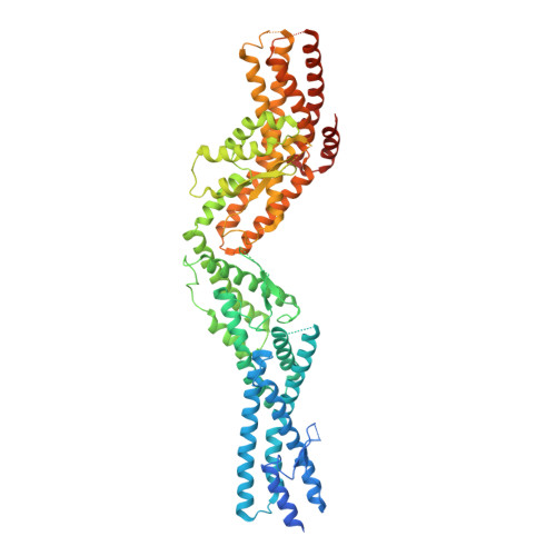
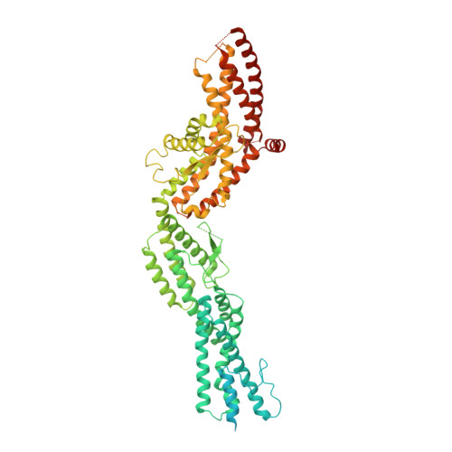
Structure of Gamma-Tubulin Small Complex Based on a Cryo-Em Map, Chemical Cross-Links, and a Remotely Related Structure.

Greenberg, C.H.Kollman, J.Zelter, A.Johnson, R.Maccoss, M.J.Davis, T.N.Agard, D.A.Sali, A.

(2016) J Struct Biol 194: 303

  • DOI: https://doi.org/10.1016/j.jsb.2016.03.006
  • Primary Citation of Related Structures:  
    5FLZ, 5FM1

  • PubMed Abstract: 

    Modeling protein complex structures based on distantly related homologues can be challenging due to poor sequence and structure conservation. Therefore, utilizing even low-resolution experimental data can significantly increase model precision and accuracy. Here, we present models of the two key functional states of the yeast γ-tubulin small complex (γTuSC): one for the low-activity "open" state and another for the higher-activity "closed" state. Both models were computed based on remotely related template structures and cryo-EM density maps at 6.9Å and 8.0Å resolution, respectively. For each state, extensive sampling of alignments and conformations was guided by the fit to the corresponding cryo-EM density map. The resulting good-scoring models formed a tightly clustered ensemble of conformations in most regions. We found significant structural differences between the two states, primarily in the γ-tubulin subunit regions where the microtubule binds. We also report a set of chemical cross-links that were found to be consistent with equilibrium between the open and closed states. The protocols developed here have been incorporated into our open-source Integrative Modeling Platform (IMP) software package (http://integrativemodeling.org), and can therefore be applied to many other systems.


  • Organizational Affiliation

    Department of Bioengineering and Therapeutic Sciences, Department of Pharmaceutical Chemistry, California Institute for Quantitative Biosciences, University of California at San Francisco, San Francisco, CA, USA. Electronic address: cgreen@salilab.org.


Macromolecules
Find similar proteins by:  (by identity cutoff)  |  3D Structure
Entity ID: 1
MoleculeChains Sequence LengthOrganismDetailsImage
SPINDLE POLE BODY COMPONENT SPC97823Saccharomyces cerevisiaeMutation(s): 0 
UniProt
Find proteins for P38863 (Saccharomyces cerevisiae (strain ATCC 204508 / S288c))
Explore P38863 
Go to UniProtKB:  P38863
Entity Groups  
Sequence Clusters30% Identity50% Identity70% Identity90% Identity95% Identity100% Identity
UniProt GroupP38863
Sequence Annotations
Expand
  • Reference Sequence
Find similar proteins by:  (by identity cutoff)  |  3D Structure
Entity ID: 2
MoleculeChains Sequence LengthOrganismDetailsImage
SPINDLE POLE BODY COMPONENT SPC98846Saccharomyces cerevisiaeMutation(s): 0 
UniProt
Find proteins for P53540 (Saccharomyces cerevisiae (strain ATCC 204508 / S288c))
Explore P53540 
Go to UniProtKB:  P53540
Entity Groups  
Sequence Clusters30% Identity50% Identity70% Identity90% Identity95% Identity100% Identity
UniProt GroupP53540
Sequence Annotations
Expand
  • Reference Sequence
Find similar proteins by:  (by identity cutoff)  |  3D Structure
Entity ID: 3
MoleculeChains Sequence LengthOrganismDetailsImage
TUBULIN GAMMA CHAIN
C, D
473Saccharomyces cerevisiaeMutation(s): 0 
UniProt
Find proteins for P53378 (Saccharomyces cerevisiae (strain ATCC 204508 / S288c))
Explore P53378 
Go to UniProtKB:  P53378
Entity Groups  
Sequence Clusters30% Identity50% Identity70% Identity90% Identity95% Identity100% Identity
UniProt GroupP53378
Sequence Annotations
Expand
  • Reference Sequence
Find similar proteins by:  (by identity cutoff)  |  3D Structure
Entity ID: 4
MoleculeChains Sequence LengthOrganismDetailsImage
SPINDLE POLE BODY COMPONENT 110
E, F
44Saccharomyces cerevisiaeMutation(s): 0 
Sequence Annotations
Expand
  • Reference Sequence
Experimental Data & Validation

Experimental Data

  • Method: ELECTRON MICROSCOPY
  • Resolution: 8.00 Å
  • Aggregation State: FILAMENT 
  • Reconstruction Method: HELICAL 
EM Software:
TaskSoftware PackageVersion
RECONSTRUCTIONSPIDER

Structure Validation

View Full Validation Report


Entry History 

Deposition Data

Revision History  (Full details and data files)

  • Version 1.0: 2016-02-03
    Type: Initial release
  • Version 1.1: 2016-03-23
    Changes: Database references
  • Version 1.2: 2016-05-11
    Changes: Database references, Other
  • Version 1.3: 2017-08-23
    Changes: Data collection