5DRO

Structure of the Aquifex aeolicus LpxC/LPC-011 Complex


Experimental Data Snapshot

  • Method: X-RAY DIFFRACTION
  • Resolution: 2.01 Å
  • R-Value Free: 0.186 
  • R-Value Work: 0.156 
  • R-Value Observed: 0.158 

wwPDB Validation   3D Report Full Report


Ligand Structure Quality Assessment 


This is version 1.3 of the entry. See complete history


Literature

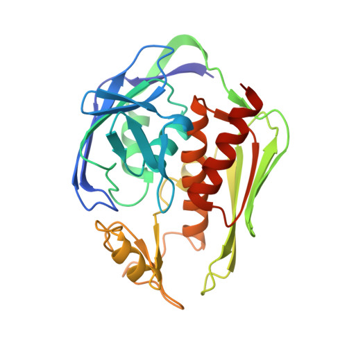
Drug design from the cryptic inhibitor envelope.

Lee, C.J.Liang, X.Wu, Q.Najeeb, J.Zhao, J.Gopalaswamy, R.Titecat, M.Sebbane, F.Lemaitre, N.Toone, E.J.Zhou, P.

(2016) Nat Commun 7: 10638-10638

  • DOI: https://doi.org/10.1038/ncomms10638
  • Primary Citation of Related Structures:  
    5DRO, 5DRP, 5DRQ, 5DRR

  • PubMed Abstract: 

    Conformational dynamics plays an important role in enzyme catalysis, allosteric regulation of protein functions and assembly of macromolecular complexes. Despite these well-established roles, such information has yet to be exploited for drug design. Here we show by nuclear magnetic resonance spectroscopy that inhibitors of LpxC--an essential enzyme of the lipid A biosynthetic pathway in Gram-negative bacteria and a validated novel antibiotic target--access alternative, minor population states in solution in addition to the ligand conformation observed in crystal structures. These conformations collectively delineate an inhibitor envelope that is invisible to crystallography, but is dynamically accessible by small molecules in solution. Drug design exploiting such a hidden inhibitor envelope has led to the development of potent antibiotics with inhibition constants in the single-digit picomolar range. The principle of the cryptic inhibitor envelope approach may be broadly applicable to other lead optimization campaigns to yield improved therapeutics.


  • Organizational Affiliation

    Department of Biochemistry, Duke University Medical Center, Durham, North Carolina 27710, USA.


Macromolecules
Find similar proteins by:  (by identity cutoff)  |  3D Structure
Entity ID: 1
MoleculeChains Sequence LengthOrganismDetailsImage
UDP-3-O-[3-hydroxymyristoyl] N-acetylglucosamine deacetylase
A, B
274Aquifex aeolicus VF5Mutation(s): 1 
Gene Names: lpxCenvAaq_1772
EC: 3.5.1
UniProt
Find proteins for O67648 (Aquifex aeolicus (strain VF5))
Explore O67648 
Go to UniProtKB:  O67648
Entity Groups  
Sequence Clusters30% Identity50% Identity70% Identity90% Identity95% Identity100% Identity
UniProt GroupO67648
Sequence Annotations
Expand
  • Reference Sequence
Small Molecules
Ligands 4 Unique
IDChains Name / Formula / InChI Key2D Diagram3D Interactions
ZH2
Query on ZH2

Download Ideal Coordinates CCD File 
D [auth A],
G [auth B]
4-[4-(4-aminophenyl)buta-1,3-diyn-1-yl]-N-[(2S,3R)-3-hydroxy-1-(hydroxyamino)-1-oxobutan-2-yl]benzamide
C21 H19 N3 O4
SNVYDNOGPYZQEV-KUHUBIRLSA-N
DMS
Query on DMS

Download Ideal Coordinates CCD File 
E [auth A],
I [auth B]
DIMETHYL SULFOXIDE
C2 H6 O S
IAZDPXIOMUYVGZ-UHFFFAOYSA-N
ZN
Query on ZN

Download Ideal Coordinates CCD File 
C [auth A],
F [auth B]
ZINC ION
Zn
PTFCDOFLOPIGGS-UHFFFAOYSA-N
ACT
Query on ACT

Download Ideal Coordinates CCD File 
H [auth B]ACETATE ION
C2 H3 O2
QTBSBXVTEAMEQO-UHFFFAOYSA-M
Experimental Data & Validation

Experimental Data

  • Method: X-RAY DIFFRACTION
  • Resolution: 2.01 Å
  • R-Value Free: 0.186 
  • R-Value Work: 0.156 
  • R-Value Observed: 0.158 
  • Space Group: P 21 21 21
Unit Cell:
Length ( Å )Angle ( ˚ )
a = 54.82α = 90
b = 74.68β = 90
c = 135.87γ = 90
Software Package:
Software NamePurpose
XSCALEdata scaling
PHASERphasing
PHENIXrefinement
PDB_EXTRACTdata extraction

Structure Validation

View Full Validation Report


Ligand Structure Quality Assessment 


Entry History & Funding Information

Deposition Data


Funding OrganizationLocationGrant Number
National Institutes of Health/National Institute Of Allergy and Infectious Diseases (NIH/NIAID)United StatesAI055588
National Institutes of Health/National Institute Of Allergy and Infectious Diseases (NIH/NIAID)United StatesAI094475

Revision History  (Full details and data files)

  • Version 1.0: 2016-03-09
    Type: Initial release
  • Version 1.1: 2017-09-20
    Changes: Author supporting evidence, Derived calculations
  • Version 1.2: 2019-12-11
    Changes: Author supporting evidence
  • Version 1.3: 2023-09-27
    Changes: Data collection, Database references, Derived calculations, Refinement description