5DD9

Menin in complex with MI-326


Experimental Data Snapshot

  • Method: X-RAY DIFFRACTION
  • Resolution: 1.62 Å
  • R-Value Free: 0.195 
  • R-Value Work: 0.162 
  • R-Value Observed: 0.163 

wwPDB Validation   3D Report Full Report


Ligand Structure Quality Assessment 


This is version 1.4 of the entry. See complete history


Literature

Rational Design of Orthogonal Multipolar Interactions with Fluorine in Protein-Ligand Complexes.

Pollock, J.Borkin, D.Lund, G.Purohit, T.Dyguda-Kazimierowicz, E.Grembecka, J.Cierpicki, T.

(2015) J Med Chem 58: 7465-7474

  • DOI: https://doi.org/10.1021/acs.jmedchem.5b00975
  • Primary Citation of Related Structures:  
    5DD9, 5DDA, 5DDB, 5DDC, 5DDD, 5DDE, 5DDF

  • PubMed Abstract: 

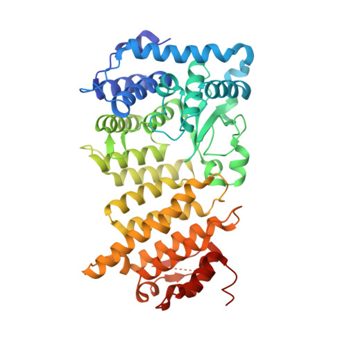
    Multipolar interactions involving fluorine and the protein backbone have been frequently observed in protein-ligand complexes. Such fluorine-backbone interactions may substantially contribute to the high affinity of small molecule inhibitors. Here we found that introduction of trifluoromethyl groups into two different sites in the thienopyrimidine class of menin-MLL inhibitors considerably improved their inhibitory activity. In both cases, trifluoromethyl groups are engaged in short interactions with the backbone of menin. In order to understand the effect of fluorine, we synthesized a series of analogues by systematically changing the number of fluorine atoms, and we determined high-resolution crystal structures of the complexes with menin. We found that introduction of fluorine at favorable geometry for interactions with backbone carbonyls may improve the activity of menin-MLL inhibitors as much as 5- to 10-fold. In order to facilitate the design of multipolar fluorine-backbone interactions in protein-ligand complexes, we developed a computational algorithm named FMAP, which calculates fluorophilic sites in proximity to the protein backbone. We demonstrated that FMAP could be used to rationalize improvement in the activity of known protein inhibitors upon introduction of fluorine. Furthermore, FMAP may also represent a valuable tool for designing new fluorine substitutions and support ligand optimization in drug discovery projects. Analysis of the menin-MLL inhibitor complexes revealed that the backbone in secondary structures is particularly accessible to the interactions with fluorine. Considering that secondary structure elements are frequently exposed at protein interfaces, we postulate that multipolar fluorine-backbone interactions may represent a particularly attractive approach to improve inhibitors of protein-protein interactions.


  • Organizational Affiliation

    Department of Pathology, University of Michigan , Ann Arbor, Michigan 48109, United States.


Macromolecules
Find similar proteins by:  (by identity cutoff)  |  3D Structure
Entity ID: 1
MoleculeChains Sequence LengthOrganismDetailsImage
Menin489Homo sapiensMutation(s): 1 
Gene Names: MEN1SCG2
UniProt & NIH Common Fund Data Resources
Find proteins for O00255 (Homo sapiens)
Explore O00255 
Go to UniProtKB:  O00255
PHAROS:  O00255
GTEx:  ENSG00000133895 
Entity Groups  
Sequence Clusters30% Identity50% Identity70% Identity90% Identity95% Identity100% Identity
UniProt GroupO00255
Sequence Annotations
Expand
  • Reference Sequence
Small Molecules
Ligands 5 Unique
IDChains Name / Formula / InChI Key2D Diagram3D Interactions
59K
Query on 59K

Download Ideal Coordinates CCD File 
B [auth A]4-[4-(5-methyl-1,3,4-thiadiazol-2-yl)piperazin-1-yl]-6-(2,2,2-trifluoroethyl)thieno[2,3-d]pyrimidine
C15 H15 F3 N6 S2
WDNNGANTDITWSR-UHFFFAOYSA-N
PG4
Query on PG4

Download Ideal Coordinates CCD File 
E [auth A],
F [auth A]
TETRAETHYLENE GLYCOL
C8 H18 O5
UWHCKJMYHZGTIT-UHFFFAOYSA-N
PGE
Query on PGE

Download Ideal Coordinates CCD File 
G [auth A]TRIETHYLENE GLYCOL
C6 H14 O4
ZIBGPFATKBEMQZ-UHFFFAOYSA-N
SO4
Query on SO4

Download Ideal Coordinates CCD File 
I [auth A],
J [auth A],
K [auth A],
L [auth A]
SULFATE ION
O4 S
QAOWNCQODCNURD-UHFFFAOYSA-L
DMS
Query on DMS

Download Ideal Coordinates CCD File 
C [auth A],
D [auth A],
H [auth A],
M [auth A],
N [auth A]
DIMETHYL SULFOXIDE
C2 H6 O S
IAZDPXIOMUYVGZ-UHFFFAOYSA-N
Binding Affinity Annotations 
IDSourceBinding Affinity
59K Binding MOAD:  5DD9 IC50: 779 (nM) from 1 assay(s)
BindingDB:  5DD9 IC50: 779 (nM) from 1 assay(s)
Experimental Data & Validation

Experimental Data

  • Method: X-RAY DIFFRACTION
  • Resolution: 1.62 Å
  • R-Value Free: 0.195 
  • R-Value Work: 0.162 
  • R-Value Observed: 0.163 
  • Space Group: P 21 21 21
Unit Cell:
Length ( Å )Angle ( ˚ )
a = 48.83α = 90
b = 80.23β = 90
c = 125.228γ = 90
Software Package:
Software NamePurpose
REFMACrefinement
HKL-2000data scaling
PDB_EXTRACTdata extraction
HKL-2000data reduction
MOLREPphasing

Structure Validation

View Full Validation Report


Ligand Structure Quality Assessment 


Entry History & Funding Information

Deposition Data


Funding OrganizationLocationGrant Number
American Cancer SocietyUnited StatesRSG-11-082-01-DMC
American Cancer SocietyUnited StatesRSG-13-130-01-CDD
National Institutes of Health/National Cancer Institute (NIH/NCI)United StatesR01 CA181185
National Institutes of Health/National Cancer Institute (NIH/NCI)United StatesR01 CA160467
Leukemia & Lymphoma SocietyUnited States6116-12
Leukemia & Lymphoma SocietyUnited States1215-14
Department of Energy (DOE, United States)United StatesDE-AC02-06CH11357
LS-CAT Sector 21United States085P1000817

Revision History  (Full details and data files)

  • Version 1.0: 2015-09-09
    Type: Initial release
  • Version 1.1: 2015-10-14
    Changes: Database references
  • Version 1.2: 2017-09-27
    Changes: Author supporting evidence, Database references, Derived calculations
  • Version 1.3: 2019-12-04
    Changes: Author supporting evidence
  • Version 1.4: 2023-09-27
    Changes: Data collection, Database references, Refinement description