5CW8

Crystal structure of Mycobacterium tuberculosis KstR in complex with 3-oxo-4-cholesten-26-oyl-CoA


Experimental Data Snapshot

  • Method: X-RAY DIFFRACTION
  • Resolution: 2.60 Å
  • R-Value Free: 0.235 
  • R-Value Work: 0.194 
  • R-Value Observed: 0.196 

wwPDB Validation   3D Report Full Report


Ligand Structure Quality Assessment 


This is version 1.4 of the entry. See complete history


Literature

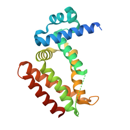
The Structure of the Transcriptional Repressor KstR in Complex with CoA Thioester Cholesterol Metabolites Sheds Light on the Regulation of Cholesterol Catabolism in Mycobacterium tuberculosis.

Ho, N.A.Dawes, S.S.Crowe, A.M.Casabon, I.Gao, C.Kendall, S.L.Baker, E.N.Eltis, L.D.Lott, J.S.

(2016) J Biol Chem 291: 7256-7266

  • DOI: https://doi.org/10.1074/jbc.M115.707760
  • Primary Citation of Related Structures:  
    3MNL, 5CW8, 5CXG, 5CXI

  • PubMed Abstract: 

    Cholesterol can be a major carbon source forMycobacterium tuberculosisduring infection, both at an early stage in the macrophage phagosome and later within the necrotic granuloma. KstR is a highly conserved TetR family transcriptional repressor that regulates a large set of genes responsible for cholesterol catabolism. Many genes in this regulon, includingkstR, are either induced during infection or are essential for survival ofM. tuberculosis in vivo In this study, we identified two ligands for KstR, both of which are CoA thioester cholesterol metabolites with four intact steroid rings. A metabolite in which one of the rings was cleaved was not a ligand. We confirmed the ligand-protein interactions using intrinsic tryptophan fluorescence and showed that ligand binding strongly inhibited KstR-DNA binding using surface plasmon resonance (IC50for ligand = 25 nm). Crystal structures of the ligand-free form of KstR show variability in the position of the DNA-binding domain. In contrast, structures of KstR·ligand complexes are highly similar to each other and demonstrate a position of the DNA-binding domain that is unfavorable for DNA binding. Comparison of ligand-bound and ligand-free structures identifies residues involved in ligand specificity and reveals a distinctive mechanism by which the ligand-induced conformational change mediates DNA release.


  • Organizational Affiliation

    From the School of Biological Sciences and Maurice Wilkins Centre for Molecular Biodiscovery, The University of Auckland, 3a Symonds Street, Auckland 1142, New Zealand.


Macromolecules
Find similar proteins by:  (by identity cutoff)  |  3D Structure
Entity ID: 1
MoleculeChains Sequence LengthOrganismDetailsImage
HTH-type transcriptional repressor KstR
A, B
203Mycobacterium tuberculosis H37RvMutation(s): 0 
Gene Names: kstRRv3574
UniProt
Find proteins for P96856 (Mycobacterium tuberculosis (strain ATCC 25618 / H37Rv))
Explore P96856 
Go to UniProtKB:  P96856
Entity Groups  
Sequence Clusters30% Identity50% Identity70% Identity90% Identity95% Identity100% Identity
UniProt GroupP96856
Sequence Annotations
Expand
  • Reference Sequence
Small Molecules
Ligands 2 Unique
IDChains Name / Formula / InChI Key2D Diagram3D Interactions
55X
Query on 55X

Download Ideal Coordinates CCD File 
C [auth A],
D [auth B]
S-{1-[5-(6-amino-9H-purin-9-yl)-4-hydroxy-3-(phosphonooxy)tetrahydrofuran-2-yl]-3,7-dihydroxy-6,6-dimethyl-3-oxido-8,12 -dioxo-2,4-dioxa-9,13-diaza-3lambda~5~-phosphapentadecan-15-yl} (2S,6R)-6-[(8S,9S,10R,13R,14S,17R)-10,13-dimethyl-3-oxo-2,3,6,7,8,9,10,11,12,13,14,15,16,17-tetradecahydro-1H-cyclopenta [a]phenanthren-17-yl]-2-methylheptanethioate (non-preferred name)
C48 H75 N7 O15 P2 S
VHFLMCCTKSGQAT-YKQRDCBHSA-N
PGE
Query on PGE

Download Ideal Coordinates CCD File 
E [auth B]TRIETHYLENE GLYCOL
C6 H14 O4
ZIBGPFATKBEMQZ-UHFFFAOYSA-N
Experimental Data & Validation

Experimental Data

  • Method: X-RAY DIFFRACTION
  • Resolution: 2.60 Å
  • R-Value Free: 0.235 
  • R-Value Work: 0.194 
  • R-Value Observed: 0.196 
  • Space Group: I 1 2 1
Unit Cell:
Length ( Å )Angle ( ˚ )
a = 49.694α = 90
b = 67.095β = 96.74
c = 124.231γ = 90
Software Package:
Software NamePurpose
PHENIXrefinement
Aimlessdata scaling
PHASERphasing
XDSdata reduction

Structure Validation

View Full Validation Report


Ligand Structure Quality Assessment 


Entry History & Funding Information

Deposition Data


Funding OrganizationLocationGrant Number
Health Research Council (HRC)New Zealand--

Revision History  (Full details and data files)

  • Version 1.0: 2016-02-17
    Type: Initial release
  • Version 1.1: 2016-02-24
    Changes: Database references
  • Version 1.2: 2016-04-20
    Changes: Database references
  • Version 1.3: 2020-01-01
    Changes: Author supporting evidence, Data collection, Database references, Derived calculations
  • Version 1.4: 2023-09-27
    Changes: Data collection, Database references, Derived calculations, Refinement description, Structure summary