5BPB

Crystal structure of the cysteine-rich domain of human Frizzled 4 - Crystal Form I


Experimental Data Snapshot

  • Method: X-RAY DIFFRACTION
  • Resolution: 2.20 Å
  • R-Value Free: 0.223 
  • R-Value Work: 0.177 
  • R-Value Observed: 0.179 

wwPDB Validation   3D Report Full Report


Ligand Structure Quality Assessment 


This is version 2.2 of the entry. See complete history


Literature

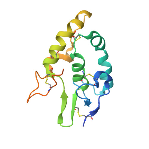
Structure and functional properties of Norrin mimic Wnt for signalling with Frizzled4, Lrp5/6, and proteoglycan.

Chang, T.H.Hsieh, F.L.Zebisch, M.Harlos, K.Elegheert, J.Jones, E.Y.

(2015) Elife 4: e06554

  • DOI: https://doi.org/10.7554/eLife.06554
  • Primary Citation of Related Structures:  
    5BPB, 5BPQ, 5BPU, 5BQ8, 5BQB, 5BQC, 5BQE

  • PubMed Abstract: 

    Wnt signalling regulates multiple processes including angiogenesis, inflammation, and tumorigenesis. Norrin (Norrie Disease Protein) is a cystine-knot like growth factor. Although unrelated to Wnt, Norrin activates the Wnt/β-catenin pathway. Signal complex formation involves Frizzled4 (Fz4), low-density lipoprotein receptor related protein 5/6 (Lrp5/6), Tetraspanin-12 and glycosaminoglycans (GAGs). Here, we report crystallographic and small-angle X-ray scattering analyses of Norrin in complex with Fz4 cysteine-rich domain (Fz4CRD), of this complex bound with GAG analogues, and of unliganded Norrin and Fz4CRD. Our structural, biophysical and cellular data, map Fz4 and putative Lrp5/6 binding sites to distinct patches on Norrin, and reveal a GAG binding site spanning Norrin and Fz4CRD. These results explain numerous disease-associated mutations. Comparison with the Xenopus Wnt8-mouse Fz8CRD complex reveals Norrin mimics Wnt for Frizzled recognition. The production and characterization of wild-type and mutant Norrins reported here open new avenues for the development of therapeutics to combat abnormal Norrin/Wnt signalling.


  • Organizational Affiliation

    Division of Structural Biology, Wellcome Trust Centre for Human Genetics, University of Oxford, Oxford, United Kingdom.


Macromolecules
Find similar proteins by:  (by identity cutoff)  |  3D Structure
Entity ID: 1
MoleculeChains Sequence LengthOrganismDetailsImage
Frizzled-4A,
B,
C [auth D],
D [auth C]
149Homo sapiensMutation(s): 0 
Gene Names: FZD4
UniProt & NIH Common Fund Data Resources
Find proteins for Q9ULV1 (Homo sapiens)
Explore Q9ULV1 
Go to UniProtKB:  Q9ULV1
PHAROS:  Q9ULV1
GTEx:  ENSG00000174804 
Entity Groups  
Sequence Clusters30% Identity50% Identity70% Identity90% Identity95% Identity100% Identity
UniProt GroupQ9ULV1
Sequence Annotations
Expand
  • Reference Sequence
Experimental Data & Validation

Experimental Data

  • Method: X-RAY DIFFRACTION
  • Resolution: 2.20 Å
  • R-Value Free: 0.223 
  • R-Value Work: 0.177 
  • R-Value Observed: 0.179 
  • Space Group: P 21 21 21
Unit Cell:
Length ( Å )Angle ( ˚ )
a = 72.608α = 90
b = 102.14β = 90
c = 116.508γ = 90
Software Package:
Software NamePurpose
PDB_EXTRACTdata extraction
XDSdata reduction
Aimlessdata scaling
PHASERphasing
Cootmodel building
REFMACrefinement

Structure Validation

View Full Validation Report


Ligand Structure Quality Assessment 


Entry History & Funding Information

Deposition Data


Funding OrganizationLocationGrant Number
Medical Research Council (United Kingdom)United KingdomG0900084
Cancer Research UKUnited KingdomC375/A10976
Wellcome TrustUnited Kingdom090532/Z/09/Z

Revision History  (Full details and data files)

  • Version 1.0: 2015-07-01
    Type: Initial release
  • Version 1.1: 2015-07-22
    Changes: Database references
  • Version 1.2: 2015-07-29
    Changes: Database references
  • Version 2.0: 2017-09-13
    Changes: Atomic model, Author supporting evidence
  • Version 2.1: 2020-07-29
    Type: Remediation
    Reason: Carbohydrate remediation
    Changes: Advisory, Data collection, Derived calculations, Structure summary
  • Version 2.2: 2024-01-10
    Changes: Data collection, Database references, Refinement description, Structure summary