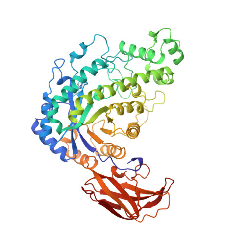
Crystal structure of beta-amylase from Bacillus cereus var. mycoides at 2.2 A resolution.
Oyama, T., Kusunoki, M., Kishimoto, Y., Takasaki, Y., Nitta, Y.(1999) J Biochem 125: 1120-1130
- PubMed: 10348915
- DOI: https://doi.org/10.1093/oxfordjournals.jbchem.a022394
- Primary Citation of Related Structures:
5BCA - PubMed Abstract:
The crystal structure of beta-amylase from Bacillus cereus var. mycoides was determined by the multiple isomorphous replacement method. The structure was refined to a final R-factor of 0.186 for 102,807 independent reflections with F/sigma(F) > or = 2.0 at 2.2 A resolution with root-mean-square deviations from ideality in bond lengths, and bond angles of 0.014 A and 3.00 degrees, respectively. The asymmetric unit comprises four molecules exhibiting a dimer-of-dimers structure. The enzyme, however, acts as a monomer in solution. The beta-amylase molecule folds into three domains; the first one is the N-terminal catalytic domain with a (beta/alpha)8 barrel, the second one is the excursion part from the first one, and the third one is the C-terminal domain with two almost anti-parallel beta-sheets. The active site cleft, including two putative catalytic residues (Glu172 and Glu367), is located on the carboxyl side of the central beta-sheet in the (beta/alpha)8 barrel, as in most amylases. The active site structure of the enzyme resembles that of soybean beta-amylase with slight differences. One calcium ion is bound per molecule far from the active site. The C-terminal domain has a fold similar to the raw starch binding domains of cyclodextrin glycosyltransferase and glucoamylase.
Organizational Affiliation:
Laboratory of Enzyme Chemistry, College of Agriculture, University of Osaka Prefecture, Sakai, Osaka, 599-8531, Japan. chicago@biochem. osakafu-u.ac.jp