5B64

A novel binding mode of MAGUK GK domain revealed by DLG GK domain in complex with KIF13B MBS domain


Experimental Data Snapshot

  • Method: X-RAY DIFFRACTION
  • Resolution: 2.70 Å
  • R-Value Free: 0.246 
  • R-Value Work: 0.187 
  • R-Value Observed: 0.190 

wwPDB Validation   3D Report Full Report


This is version 1.2 of the entry. See complete history


Literature

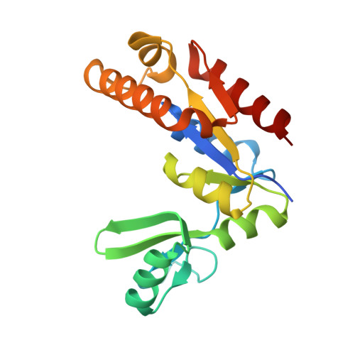
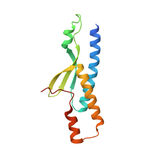
An Atypical MAGUK GK Target Recognition Mode Revealed by the Interaction between DLG and KIF13B

Zhu, J.Shang, Y.Xia, Y.Zhang, R.Zhang, M.

(2016) Structure 24: 1876-1885

  • DOI: https://doi.org/10.1016/j.str.2016.08.008
  • Primary Citation of Related Structures:  
    5B64

  • PubMed Abstract: 

    The membrane-associated guanylate kinase (MAGUK) scaffold proteins share a signature guanylate kinase (GK) domain. Despite their diverse functional roles in cell polarity control and synaptic signaling, the currently known mode of action of MAGUK GK is via its binding to phosphorylated short peptides from target proteins. Here, we discover that the GK domain of DLG MAGUK binds to an unphosphorylated and autonomously folded domain within the stalk region (MAGUK binding stalk [MBS] domain) of a kinesin motor KIF13B with high specificity and affinity. The structure of DLG4 GK in complex with KIF13B MBS reveals the molecular mechanism governing this atypical GK/target recognition mode and provides insights into DLG/KIF13B complex-mediated regulation of diverse cellular processes such as asymmetric cell division. We further show that binding to non-phosphorylated targets is another general property of MAGUK GKs, thus expanding the mechanisms of action of the MAGUK family proteins.


  • Organizational Affiliation

    National Center for Protein Science Shanghai, Institute of Biochemistry and Cell Biology, Shanghai Institutes for Biological Sciences, Chinese Academy of Sciences, Shanghai 201210, China; Division of Life Science, State Key Laboratory of Molecular Neuroscience, Hong Kong University of Science and Technology, Clear Water Bay, Kowloon, Hong Kong, China. Electronic address: jinwei.zhu@sibcb.ac.cn.


Macromolecules
Find similar proteins by:  (by identity cutoff)  |  3D Structure
Entity ID: 1
MoleculeChains Sequence LengthOrganismDetailsImage
DLG GK189RattusMutation(s): 0 
UniProt
Find proteins for P31016 (Rattus norvegicus)
Explore P31016 
Go to UniProtKB:  P31016
Entity Groups  
Sequence Clusters30% Identity50% Identity70% Identity90% Identity95% Identity100% Identity
UniProt GroupP31016
Sequence Annotations
Expand
  • Reference Sequence
Find similar proteins by:  (by identity cutoff)  |  3D Structure
Entity ID: 2
MoleculeChains Sequence LengthOrganismDetailsImage
Protein Kif13b126Mus musculusMutation(s): 0 
Gene Names: Kif13b
UniProt & NIH Common Fund Data Resources
Find proteins for E9Q4K7 (Mus musculus)
Explore E9Q4K7 
Go to UniProtKB:  E9Q4K7
IMPC:  MGI:1098265
Entity Groups  
Sequence Clusters30% Identity50% Identity70% Identity90% Identity95% Identity100% Identity
UniProt GroupE9Q4K7
Sequence Annotations
Expand
  • Reference Sequence
Experimental Data & Validation

Experimental Data

  • Method: X-RAY DIFFRACTION
  • Resolution: 2.70 Å
  • R-Value Free: 0.246 
  • R-Value Work: 0.187 
  • R-Value Observed: 0.190 
  • Space Group: I 4
Unit Cell:
Length ( Å )Angle ( ˚ )
a = 116.279α = 90
b = 116.279β = 90
c = 72.029γ = 90
Software Package:
Software NamePurpose
PHENIXrefinement
HKL-2000data reduction
HKL-2000data scaling
PHASERphasing

Structure Validation

View Full Validation Report


Entry History 

Deposition Data

Revision History  (Full details and data files)

  • Version 1.0: 2016-10-12
    Type: Initial release
  • Version 1.1: 2016-11-16
    Changes: Database references
  • Version 1.2: 2024-03-20
    Changes: Data collection, Database references, Derived calculations