5A7U

Single-particle cryo-EM of co-translational folded adr1 domain inside the E. coli ribosome exit tunnel.


Experimental Data Snapshot

  • Method: ELECTRON MICROSCOPY
  • Resolution: 4.80 Å
  • Aggregation State: PARTICLE 
  • Reconstruction Method: SINGLE PARTICLE 

wwPDB Validation   3D Report Full Report


This is version 1.2 of the entry. See complete history


Literature

Cotranslational Protein Folding Inside the Ribosome Exit Tunnel.

Nilsson, O.B.Hedman, R.Marino, J.Wickles, S.Bischoff, L.Johansson, M.Muller-Lucks, A.Trovato, F.Puglisi, J.D.O'Brien, E.P.Beckmann, R.Von Heijne, G.

(2015) Cell Rep 12: 1533

  • DOI: https://doi.org/10.1016/j.celrep.2015.07.065
  • Primary Citation of Related Structures:  
    5A7U

  • PubMed Abstract: 

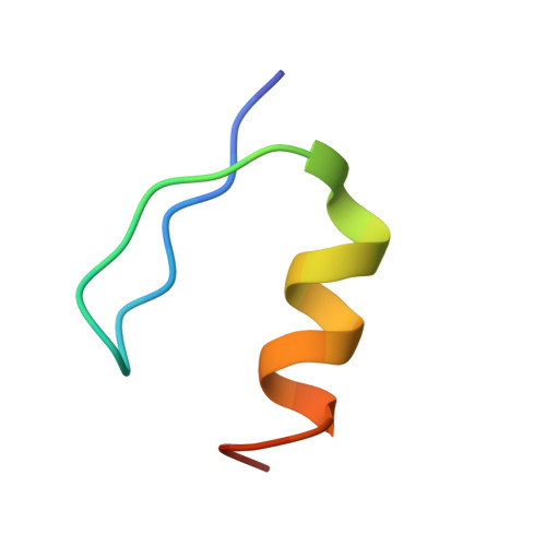
    At what point during translation do proteins fold? It is well established that proteins can fold cotranslationally outside the ribosome exit tunnel, whereas studies of folding inside the exit tunnel have so far detected only the formation of helical secondary structure and collapsed or partially structured folding intermediates. Here, using a combination of cotranslational nascent chain force measurements, inter-subunit fluorescence resonance energy transfer studies on single translating ribosomes, molecular dynamics simulations, and cryoelectron microscopy, we show that a small zinc-finger domain protein can fold deep inside the vestibule of the ribosome exit tunnel. Thus, for small protein domains, the ribosome itself can provide the kind of sheltered folding environment that chaperones provide for larger proteins.


  • Organizational Affiliation

    Department of Biochemistry and Biophysics, Center for Biomembrane Research, Stockholm University, 106 91 Stockholm, Sweden.


Macromolecules
Find similar proteins by:  (by identity cutoff)  |  3D Structure
Entity ID: 1
MoleculeChains Sequence LengthOrganismDetailsImage
REGULATORY PROTEIN ADR129Saccharomyces cerevisiaeMutation(s): 0 
UniProt
Find proteins for P07248 (Saccharomyces cerevisiae (strain ATCC 204508 / S288c))
Explore P07248 
Go to UniProtKB:  P07248
Entity Groups  
Sequence Clusters30% Identity50% Identity70% Identity90% Identity95% Identity100% Identity
UniProt GroupP07248
Sequence Annotations
Expand
  • Reference Sequence
Small Molecules
Ligands 1 Unique
IDChains Name / Formula / InChI Key2D Diagram3D Interactions
ZN
Query on ZN

Download Ideal Coordinates CCD File 
B [auth A]ZINC ION
Zn
PTFCDOFLOPIGGS-UHFFFAOYSA-N
Experimental Data & Validation

Experimental Data

  • Method: ELECTRON MICROSCOPY
  • Resolution: 4.80 Å
  • Aggregation State: PARTICLE 
  • Reconstruction Method: SINGLE PARTICLE 
EM Software:
TaskSoftware PackageVersion
RECONSTRUCTIONSPIDER

Structure Validation

View Full Validation Report


Entry History 

Revision History  (Full details and data files)

  • Version 1.0: 2015-09-16
    Type: Initial release
  • Version 1.1: 2015-09-23
    Changes: Database references
  • Version 1.2: 2017-08-23
    Changes: Data collection