5A6C

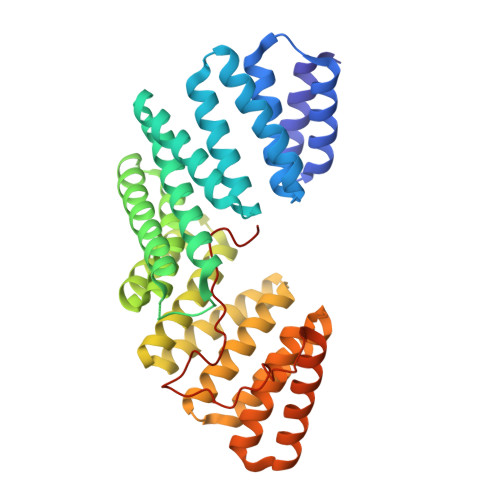
Concomitant binding of Afadin to LGN and F-actin directs planar spindle orientation


Experimental Data Snapshot

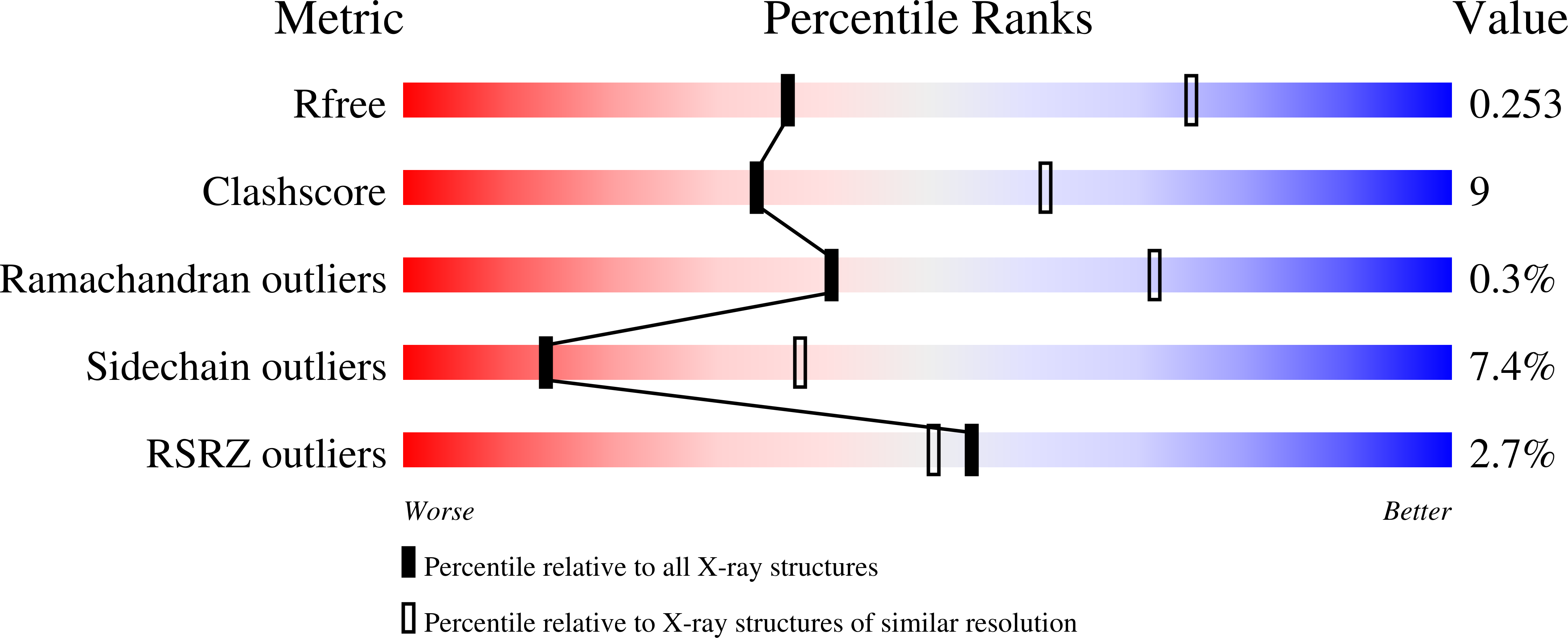
  • Method: X-RAY DIFFRACTION
  • Resolution: 2.90 Å
  • R-Value Free: 0.252 
  • R-Value Work: 0.206 
  • R-Value Observed: 0.208 

wwPDB Validation   3D Report Full Report


This is version 1.4 of the entry. See complete history


Literature

Concomitant Binding of Afadin to Lgn and F-Actin Directs Planar Spindle Orientation.

Carminati, M.Gallini, S.Pirovano, L.Alfieri, A.Bisi, S.Mapelli, M.

(2016) Nat Struct Mol Biol 23: 155

  • DOI: https://doi.org/10.1038/nsmb.3152
  • Primary Citation of Related Structures:  
    5A6C

  • PubMed Abstract: 

    Polarized epithelia form by oriented cell divisions in which the mitotic spindle aligns parallel to the epithelial plane. To orient the mitotic spindle, cortical cues trigger the recruitment of NuMA-dynein-based motors, which pull on astral microtubules via the protein LGN. We demonstrate that the junctional protein Afadin is required for spindle orientation and correct epithelial morphogenesis of Caco-2 cysts. Molecularly, Afadin binds directly and concomitantly to F-actin and to LGN. We determined the crystallographic structure of human Afadin in complex with LGN and show that it resembles the LGN-NuMA complex. In mitosis, Afadin is necessary for cortical accumulation of LGN and NuMA above the spindle poles, in an F-actin-dependent manner. Collectively, our results depict Afadin as a molecular hub governing the enrichment of LGN and NuMA at the cortex. To our knowledge, Afadin is the first-described mechanical anchor between dynein and cortical F-actin.


  • Organizational Affiliation

    Department of Experimental Oncology, European Institute of Oncology, Milan, Italy.


Macromolecules
Find similar proteins by:  (by identity cutoff)  |  3D Structure
Entity ID: 1
MoleculeChains Sequence LengthOrganismDetailsImage
G-PROTEIN-SIGNALING MODULATOR 2, AFADIN
A, B
374Homo sapiensMutation(s): 0 
Gene Names: GPSM2LGNMLLT4AF6
UniProt & NIH Common Fund Data Resources
Find proteins for P55196 (Homo sapiens)
Explore P55196 
Go to UniProtKB:  P55196
PHAROS:  P55196
GTEx:  ENSG00000130396 
Find proteins for P81274 (Homo sapiens)
Explore P81274 
Go to UniProtKB:  P81274
PHAROS:  P81274
GTEx:  ENSG00000121957 
Entity Groups  
Sequence Clusters30% Identity50% Identity70% Identity90% Identity95% Identity100% Identity
UniProt GroupsP55196P81274
Sequence Annotations
Expand
  • Reference Sequence
Experimental Data & Validation

Experimental Data

  • Method: X-RAY DIFFRACTION
  • Resolution: 2.90 Å
  • R-Value Free: 0.252 
  • R-Value Work: 0.206 
  • R-Value Observed: 0.208 
  • Space Group: P 21 3
Unit Cell:
Length ( Å )Angle ( ˚ )
a = 169.88α = 90
b = 169.88β = 90
c = 169.88γ = 90
Software Package:
Software NamePurpose
PHENIXrefinement
XDSdata reduction
SCALAdata scaling
PHASERphasing

Structure Validation

View Full Validation Report


Entry History 

Deposition Data

Revision History  (Full details and data files)

  • Version 1.0: 2015-12-30
    Type: Initial release
  • Version 1.1: 2016-01-20
    Changes: Database references
  • Version 1.2: 2016-01-27
    Changes: Structure summary
  • Version 1.3: 2016-03-02
    Changes: Database references
  • Version 1.4: 2024-01-10
    Changes: Data collection, Database references, Derived calculations, Other, Refinement description