5NAL

The crystal structure of inhibitor-15 covalently bound to PDE6D


Experimental Data Snapshot

  • Method: X-RAY DIFFRACTION
  • Resolution: 2.20 Å
  • R-Value Free: 0.243 
  • R-Value Work: 0.195 
  • R-Value Observed: 0.198 

wwPDB Validation   3D Report Full Report


Ligand Structure Quality Assessment 


This is version 1.2 of the entry. See complete history


Literature

Covalent Protein Labeling at Glutamic Acids.

Martin-Gago, P.Fansa, E.K.Winzker, M.Murarka, S.Janning, P.Schultz-Fademrecht, C.Baumann, M.Wittinghofer, A.Waldmann, H.

(2017) Cell Chem Biol 24: 589-597.e5

  • DOI: https://doi.org/10.1016/j.chembiol.2017.03.015
  • Primary Citation of Related Structures:  
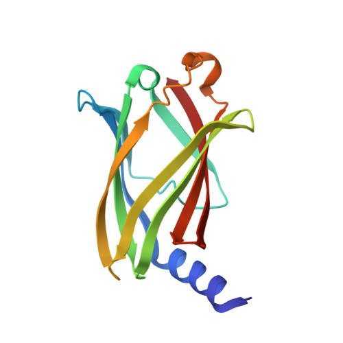
    5NAL

  • PubMed Abstract: 

    Covalent labeling of amino acids in proteins by reactive small molecules, in particular at cysteine SH and lysine NH groups, is a powerful approach to identify and characterize proteins and their functions. However, for the less-reactive carboxylic acids present in Asp and Glu, hardly any methodology is available. Employing the lipoprotein binding chaperone PDE6δ as an example, we demonstrate that incorporation of isoxazolium salts that resemble the structure and reactivity of Woodward's reagent K into protein ligands provides a novel method for selective covalent targeting of binding site carboxylic acids in whole proteomes. Covalent adduct formation occurs via rapid formation of enol esters and the covalent bond is stable even in the presence of strong nucleophiles. This new method promises to open up hitherto unexplored opportunities for chemical biology research.


  • Organizational Affiliation

    Department of Chemical Biology, Max Planck Institute of Molecular Physiology, Otto-Hahn Straße 11, 44227 Dortmund, Germany.


Macromolecules
Find similar proteins by:  (by identity cutoff)  |  3D Structure
Entity ID: 1
MoleculeChains Sequence LengthOrganismDetailsImage
Retinal rod rhodopsin-sensitive cGMP 3',5'-cyclic phosphodiesterase subunit deltaA [auth B]150Homo sapiensMutation(s): 0 
Gene Names: PDE6DPDED
UniProt & NIH Common Fund Data Resources
Find proteins for O43924 (Homo sapiens)
Explore O43924 
Go to UniProtKB:  O43924
PHAROS:  O43924
GTEx:  ENSG00000156973 
Entity Groups  
Sequence Clusters30% Identity50% Identity70% Identity90% Identity95% Identity100% Identity
UniProt GroupO43924
Sequence Annotations
Expand
  • Reference Sequence
Small Molecules
Ligands 1 Unique
IDChains Name / Formula / InChI Key2D Diagram3D Interactions
8RQ
Query on 8RQ

Download Ideal Coordinates CCD File 
B
~{N}4-[(4-chlorophenyl)methyl]-~{N}1-(cyclohexylmethyl)-~{N}4-cyclopentyl-~{N}1-[(~{Z})-4-[(~{E})-methyliminomethyl]-5-oxidanyl-hex-4-enyl]benzene-1,4-disulfonamide
C33 H46 Cl N3 O5 S2
BJGXSIOLQNSMNV-ATLOAHSKSA-N
Experimental Data & Validation

Experimental Data

  • Method: X-RAY DIFFRACTION
  • Resolution: 2.20 Å
  • R-Value Free: 0.243 
  • R-Value Work: 0.195 
  • R-Value Observed: 0.198 
  • Space Group: P 32 2 1
Unit Cell:
Length ( Å )Angle ( ˚ )
a = 56.07α = 90
b = 56.07β = 90
c = 115.08γ = 120
Software Package:
Software NamePurpose
XSCALEdata scaling
REFMACrefinement
PDB_EXTRACTdata extraction
XDSdata reduction
MOLREPphasing

Structure Validation

View Full Validation Report


Ligand Structure Quality Assessment 


Entry History 

Deposition Data

Revision History  (Full details and data files)

  • Version 1.0: 2017-05-10
    Type: Initial release
  • Version 1.1: 2017-05-31
    Changes: Database references
  • Version 1.2: 2024-01-17
    Changes: Data collection, Database references, Refinement description