5ICN

HDAC1:MTA1 in complex with inositol-6-phosphate and a novel peptide inhibitor based on histone H4


Experimental Data Snapshot

  • Method: X-RAY DIFFRACTION
  • Resolution: 3.30 Å
  • R-Value Free: 0.299 
  • R-Value Work: 0.255 
  • R-Value Observed: 0.257 

wwPDB Validation   3D Report Full Report


Ligand Structure Quality Assessment 


This is version 2.1 of the entry. See complete history


Literature

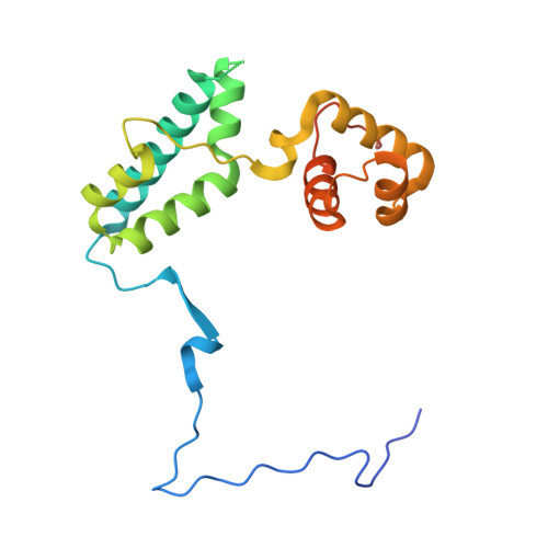
Insights into the activation mechanism of class I HDAC complexes by inositol phosphates.

Watson, P.J.Millard, C.J.Riley, A.M.Robertson, N.S.Wright, L.C.Godage, H.Y.Cowley, S.M.Jamieson, A.G.Potter, B.V.Schwabe, J.W.

(2016) Nat Commun 7: 11262-11262

  • DOI: https://doi.org/10.1038/ncomms11262
  • Primary Citation of Related Structures:  
    5ICN

  • PubMed Abstract: 

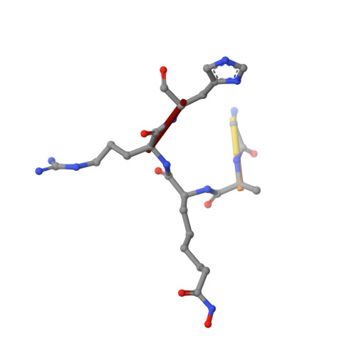
    Histone deacetylases (HDACs) 1, 2 and 3 form the catalytic subunit of several large transcriptional repression complexes. Unexpectedly, the enzymatic activity of HDACs in these complexes has been shown to be regulated by inositol phosphates, which bind in a pocket sandwiched between the HDAC and co-repressor proteins. However, the actual mechanism of activation remains poorly understood. Here we have elucidated the stereochemical requirements for binding and activation by inositol phosphates, demonstrating that activation requires three adjacent phosphate groups and that other positions on the inositol ring can tolerate bulky substituents. We also demonstrate that there is allosteric communication between the inositol-binding site and the active site. The crystal structure of the HDAC1:MTA1 complex bound to a novel peptide-based inhibitor and to inositol hexaphosphate suggests a molecular basis of substrate recognition, and an entropically driven allosteric mechanism of activation.


  • Organizational Affiliation

    Henry Wellcome Laboratories of Structural Biology, Department of Molecular and Cell Biology, University of Leicester, Leicester LE1 9HN, UK.


Macromolecules
Find similar proteins by:  (by identity cutoff)  |  3D Structure
Entity ID: 1
MoleculeChains Sequence LengthOrganismDetailsImage
Metastasis-associated protein MTA1195Homo sapiensMutation(s): 0 
Gene Names: MTA1
UniProt & NIH Common Fund Data Resources
Find proteins for Q13330 (Homo sapiens)
Explore Q13330 
Go to UniProtKB:  Q13330
PHAROS:  Q13330
GTEx:  ENSG00000182979 
Entity Groups  
Sequence Clusters30% Identity50% Identity70% Identity90% Identity95% Identity100% Identity
UniProt GroupQ13330
Sequence Annotations
Expand
  • Reference Sequence
Find similar proteins by:  (by identity cutoff)  |  3D Structure
Entity ID: 2
MoleculeChains Sequence LengthOrganismDetailsImage
Histone deacetylase 1376Homo sapiensMutation(s): 0 
Gene Names: HDAC1RPD3L1
EC: 3.5.1.98
UniProt & NIH Common Fund Data Resources
Find proteins for Q13547 (Homo sapiens)
Explore Q13547 
Go to UniProtKB:  Q13547
PHAROS:  Q13547
GTEx:  ENSG00000116478 
Entity Groups  
Sequence Clusters30% Identity50% Identity70% Identity90% Identity95% Identity100% Identity
UniProt GroupQ13547
Sequence Annotations
Expand
  • Reference Sequence

Find similar proteins by:  Sequence   |   3D Structure  

Entity ID: 3
MoleculeChains Sequence LengthOrganismDetailsImage
GLY-ALA-6A0-ARG-HIS9synthetic constructMutation(s): 0 
Sequence Annotations
Expand
  • Reference Sequence
Experimental Data & Validation

Experimental Data

  • Method: X-RAY DIFFRACTION
  • Resolution: 3.30 Å
  • R-Value Free: 0.299 
  • R-Value Work: 0.255 
  • R-Value Observed: 0.257 
  • Space Group: P 32 2 1
Unit Cell:
Length ( Å )Angle ( ˚ )
a = 107.815α = 90
b = 107.815β = 90
c = 134.161γ = 120
Software Package:
Software NamePurpose
REFMACrefinement
iMOSFLMdata reduction
Aimlessdata scaling
PHASERphasing

Structure Validation

View Full Validation Report


Ligand Structure Quality Assessment 


Entry History & Funding Information

Deposition Data


Funding OrganizationLocationGrant Number
Wellcome TrustUnited KingdomWT085408
Wellcome TrustUnited KingdomWT100237
Wellcome TrustUnited KingdomWT082837
Wellcome TrustUnited KingdomWT101010
Wolfson FoundationUnited Kingdom--

Revision History  (Full details and data files)

  • Version 1.0: 2016-05-11
    Type: Initial release
  • Version 1.1: 2017-09-13
    Changes: Author supporting evidence
  • Version 1.2: 2018-01-24
    Changes: Source and taxonomy
  • Version 2.0: 2020-10-07
    Changes: Atomic model, Data collection, Derived calculations, Non-polymer description, Structure summary
  • Version 2.1: 2024-01-10
    Changes: Data collection, Database references, Derived calculations, Refinement description