5EO9

Crystal Structure of the complex of Dpr6 Domain 1 bound to DIP-alpha Domain 1+2


Experimental Data Snapshot

  • Method: X-RAY DIFFRACTION
  • Resolution: 2.30 Å
  • R-Value Free: 0.252 
  • R-Value Work: 0.202 
  • R-Value Observed: 0.205 

wwPDB Validation   3D Report Full Report


Ligand Structure Quality Assessment 


This is version 1.2 of the entry. See complete history


Literature

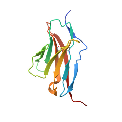
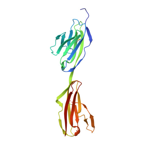
Control of Synaptic Connectivity by a Network of Drosophila IgSF Cell Surface Proteins.

Carrillo, R.A.Ozkan, E.Menon, K.P.Nagarkar-Jaiswal, S.Lee, P.T.Jeon, M.Birnbaum, M.E.Bellen, H.J.Garcia, K.C.Zinn, K.

(2015) Cell 163: 1770-1782

  • DOI: https://doi.org/10.1016/j.cell.2015.11.022
  • Primary Citation of Related Structures:  
    5EO9

  • PubMed Abstract: 

    We have defined a network of interacting Drosophila cell surface proteins in which a 21-member IgSF subfamily, the Dprs, binds to a nine-member subfamily, the DIPs. The structural basis of the Dpr-DIP interaction code appears to be dictated by shape complementarity within the Dpr-DIP binding interface. Each of the six dpr and DIP genes examined here is expressed by a unique subset of larval and pupal neurons. In the neuromuscular system, interactions between Dpr11 and DIP-γ affect presynaptic terminal development, trophic factor responses, and neurotransmission. In the visual system, dpr11 is selectively expressed by R7 photoreceptors that use Rh4 opsin (yR7s). Their primary synaptic targets, Dm8 amacrine neurons, express DIP-γ. In dpr11 or DIP-γ mutants, yR7 terminals extend beyond their normal termination zones in layer M6 of the medulla. DIP-γ is also required for Dm8 survival or differentiation. Our findings suggest that Dpr-DIP interactions are important determinants of synaptic connectivity.


  • Organizational Affiliation

    Division of Biology and Biological Engineering, California Institute of Technology, Pasadena, CA.


Macromolecules
Find similar proteins by:  (by identity cutoff)  |  3D Structure
Entity ID: 1
MoleculeChains Sequence LengthOrganismDetailsImage
Dpr6, isoform C110Drosophila melanogasterMutation(s): 0 
Gene Names: dpr6CG14162Dmel_CG14162
UniProt
Find proteins for M9PC40 (Drosophila melanogaster)
Explore M9PC40 
Go to UniProtKB:  M9PC40
Entity Groups  
Sequence Clusters30% Identity50% Identity70% Identity90% Identity95% Identity100% Identity
UniProt GroupM9PC40
Sequence Annotations
Expand
  • Reference Sequence
Find similar proteins by:  (by identity cutoff)  |  3D Structure
Entity ID: 2
MoleculeChains Sequence LengthOrganismDetailsImage
CG32791, isoform A206Drosophila melanogasterMutation(s): 0 
Gene Names: DIP-alphaCG13020CG32791Dmel_CG32791
UniProt
Find proteins for Q9W4R3 (Drosophila melanogaster)
Explore Q9W4R3 
Go to UniProtKB:  Q9W4R3
Entity Groups  
Sequence Clusters30% Identity50% Identity70% Identity90% Identity95% Identity100% Identity
UniProt GroupQ9W4R3
Sequence Annotations
Expand
  • Reference Sequence
Experimental Data & Validation

Experimental Data

Unit Cell:
Length ( Å )Angle ( ˚ )
a = 130.247α = 90
b = 49.299β = 90
c = 54.707γ = 90
Software Package:
Software NamePurpose
PHENIXrefinement
HKL-2000data reduction
HKL-2000data scaling
autoSHARPphasing
SHELXphasing

Structure Validation

View Full Validation Report


Ligand Structure Quality Assessment 


Entry History & Funding Information

Deposition Data


Funding OrganizationLocationGrant Number
Howard Hughes Medical Institute (HHMI)United States--

Revision History  (Full details and data files)

  • Version 1.0: 2016-01-06
    Type: Initial release
  • Version 1.1: 2019-11-20
    Changes: Author supporting evidence, Derived calculations
  • Version 1.2: 2020-07-29
    Type: Remediation
    Reason: Carbohydrate remediation
    Changes: Data collection, Derived calculations, Structure summary