Crystal Structure of the Apo-Form of NADPH-Dependent Thioredoxin Reductase from a Methane-Producing Archaeon.
Buey, R.M., Schmitz, R.A., Buchanan, B.B., Balsera, M.(2018) Antioxidants (Basel) 7
- PubMed: 30453601
- DOI: https://doi.org/10.3390/antiox7110166
- Primary Citation of Related Structures:
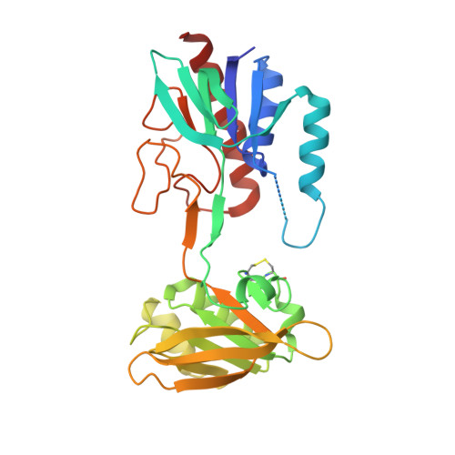
4ZN0 - PubMed Abstract:
The redox regulation of proteins via reversible dithiol/disulfide exchange reactions involves the thioredoxin system, which is composed of a reductant, a thioredoxin reductase (TR), and thioredoxin (Trx). In the pyridine nucleotide-dependent Trx reduction pathway, reducing equivalents, typically from reduced nicotinamide adenine dinucleotide phosphate (NADPH), are transferred from NADPH-TR (NTR) to Trx and, in turn, to target proteins, thus resulting in the reversible modification of the structural and functional properties of the targets. NTR enzymes contain three functional sites: an NADPH binding pocket, a non-covalently bound flavin cofactor, and a redox-active disulfide in the form of CxxC. With the aim of increasing our knowledge of the thioredoxin system in archaea, we here report the high-resolution crystal structure of NTR from the methane-generating organism Methanosarcina mazei strain Gö1 (MmNTR) at 2.6 Å resolution. Based on the crystals presently described, MmNTR assumes an overall fold that is nearly identical to the archetypal fold of authentic NTRs; however, surprisingly, we observed no electron density for flavin adenine dinucleotide (FAD) despite the well-defined and conserved FAD-binding cavity in the folded module. Remarkably, the dimers of the apo-protein within the crystal were different from those observed by small angle X-ray scattering (SAXS) for the holo-protein, suggesting that the binding of the flavin cofactor does not require major protein structural rearrangements. Rather, binding results in the stabilization of essential parts of the structure, such as those involved in dimer stabilization. Altogether, this structure represents the example of an apo-form of an NTR that yields important insight into the effects of the cofactor on protein folding.
- Metabolic Engineering Group. Dpto. Microbiología y Genética. Universidad de Salamanca, 37007 Salamanca, Spain. ruben.martinez@usal.es.
Organizational Affiliation: