4ZBX

Family 4 uracil-DNA glycosylase from Sulfolobus tokodaii (free form, X-ray wavelength=0.9000)


Experimental Data Snapshot

  • Method: X-RAY DIFFRACTION
  • Resolution: 1.70 Å
  • R-Value Free: 0.216 
  • R-Value Work: 0.169 
  • R-Value Observed: 0.171 

wwPDB Validation   3D Report Full Report


This is version 1.4 of the entry. See complete history


Literature

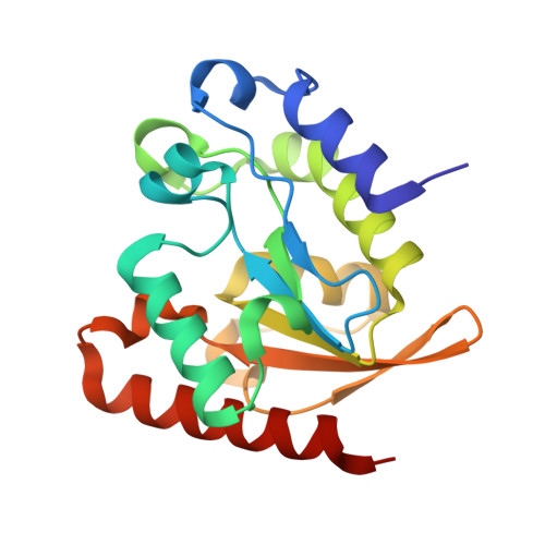
Crystal structure of family 4 uracil-DNA glycosylase from Sulfolobus tokodaii and a function of tyrosine 170 in DNA binding

Kawai, A.Higuchi, S.Tsunoda, M.Nakamura, K.T.Yamagata, Y.Miyamoto, S.

(2015) FEBS Lett 589: 2675-2682

  • DOI: https://doi.org/10.1016/j.febslet.2015.08.019
  • Primary Citation of Related Structures:  
    4ZBX, 4ZBY, 4ZBZ

  • PubMed Abstract: 

    Uracil-DNA glycosylases (UDGs) excise uracil from DNA by catalyzing the N-glycosidic bond hydrolysis. Here we report the first crystal structures of an archaeal UDG (stoUDG). Compared with other UDGs, stoUDG has a different structure of the leucine-intercalation loop, which is important for DNA binding. The stoUDG-DNA complex model indicated that Leu169, Tyr170, and Asn171 in the loop are involved in DNA intercalation. Mutational analysis showed that Tyr170 is critical for substrate DNA recognition. These results indicate that Tyr170 occupies the intercalation site formed after the structural change of the leucine-intercalation loop required for the catalysis.


  • Organizational Affiliation

    Faculty of Pharmaceutical Sciences, Sojo University, 4-22-1 Ikeda, Nishi-ku, Kumamoto 860-0082, Japan. Electronic address: akawai@ph.sojo-u.ac.jp.


Macromolecules
Find similar proteins by:  (by identity cutoff)  |  3D Structure
Entity ID: 1
MoleculeChains Sequence LengthOrganismDetailsImage
Uracil-DNA glycosylase194Sulfurisphaera tokodaii str. 7Mutation(s): 0 
Gene Names: STK_22380
EC: 3.2.2.27
UniProt
Find proteins for Q96YD0 (Sulfurisphaera tokodaii (strain DSM 16993 / JCM 10545 / NBRC 100140 / 7))
Explore Q96YD0 
Go to UniProtKB:  Q96YD0
Entity Groups  
Sequence Clusters30% Identity50% Identity70% Identity90% Identity95% Identity100% Identity
UniProt GroupQ96YD0
Sequence Annotations
Expand
  • Reference Sequence
Small Molecules
Ligands 2 Unique
IDChains Name / Formula / InChI Key2D Diagram3D Interactions
SF4
Query on SF4

Download Ideal Coordinates CCD File 
B [auth A]IRON/SULFUR CLUSTER
Fe4 S4
LJBDFODJNLIPKO-UHFFFAOYSA-N
MES
Query on MES

Download Ideal Coordinates CCD File 
C [auth A]2-(N-MORPHOLINO)-ETHANESULFONIC ACID
C6 H13 N O4 S
SXGZJKUKBWWHRA-UHFFFAOYSA-N
Experimental Data & Validation

Experimental Data

  • Method: X-RAY DIFFRACTION
  • Resolution: 1.70 Å
  • R-Value Free: 0.216 
  • R-Value Work: 0.169 
  • R-Value Observed: 0.171 
  • Space Group: P 21 21 21
Unit Cell:
Length ( Å )Angle ( ˚ )
a = 52.189α = 90
b = 52.25β = 90
c = 74.453γ = 90
Software Package:
Software NamePurpose
PHENIXrefinement
HKL-2000data processing
HKL-2000data scaling
MOLREPphasing

Structure Validation

View Full Validation Report


Entry History 

Deposition Data

Revision History  (Full details and data files)

  • Version 1.0: 2015-09-09
    Type: Initial release
  • Version 1.1: 2015-09-16
    Changes: Database references
  • Version 1.2: 2015-09-23
    Changes: Database references
  • Version 1.3: 2020-02-05
    Changes: Data collection, Database references, Derived calculations
  • Version 1.4: 2023-11-08
    Changes: Data collection, Database references, Refinement description