4Z07

Co-crystal structure of the tandem CNB (CNB-A/B) domains of human PKG I beta with cGMP


Experimental Data Snapshot

  • Method: X-RAY DIFFRACTION
  • Resolution: 2.50 Å
  • R-Value Free: 0.224 
  • R-Value Work: 0.162 
  • R-Value Observed: 0.165 

wwPDB Validation   3D Report Full Report


Ligand Structure Quality Assessment 


This is version 1.6 of the entry. See complete history


Literature

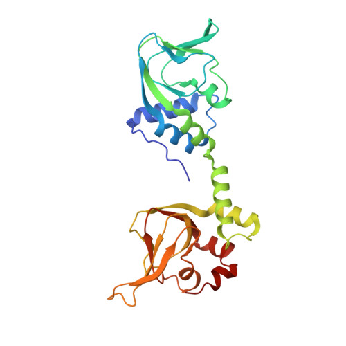
Crystal Structure of PKG I:cGMP Complex Reveals a cGMP-Mediated Dimeric Interface that Facilitates cGMP-Induced Activation.

Kim, J.J.Lorenz, R.Arold, S.T.Reger, A.S.Sankaran, B.Casteel, D.E.Herberg, F.W.Kim, C.

(2016) Structure 24: 710-720

  • DOI: https://doi.org/10.1016/j.str.2016.03.009
  • Primary Citation of Related Structures:  
    4Z07

  • PubMed Abstract: 

    Cyclic guanosine monophosphate (cGMP)-dependent protein kinase (PKG) is a key regulator of smooth muscle and vascular tone and represents an important drug target for treating hypertensive diseases and erectile dysfunction. Despite its importance, its activation mechanism is not fully understood. To understand the activation mechanism, we determined a 2.5 Å crystal structure of the PKG I regulatory (R) domain bound with cGMP, which represents the activated state. Although we used a monomeric domain for crystallization, the structure reveals that two R domains form a symmetric dimer where the cGMP bound at high-affinity pockets provide critical dimeric contacts. Small-angle X-ray scattering and mutagenesis support this dimer model, suggesting that the dimer interface modulates kinase activation. Finally, structural comparison with the homologous cyclic AMP-dependent protein kinase reveals that PKG is drastically different from protein kinase A in its active conformation, suggesting a novel activation mechanism for PKG.


  • Organizational Affiliation

    Department of Pharmacology, Baylor College of Medicine, Houston, TX 77030, USA; Department of Biochemistry, University of Kassel, Kassel, Hesse 34132, Germany.


Macromolecules
Find similar proteins by:  (by identity cutoff)  |  3D Structure
Entity ID: 1
MoleculeChains Sequence LengthOrganismDetailsImage
cGMP-dependent protein kinase 1A,
B [auth C],
C [auth E]
262Homo sapiensMutation(s): 0 
Gene Names: PRKG1PRKG1BPRKGR1APRKGR1B
EC: 2.7.11.12
UniProt & NIH Common Fund Data Resources
Find proteins for Q13976 (Homo sapiens)
Explore Q13976 
Go to UniProtKB:  Q13976
PHAROS:  Q13976
GTEx:  ENSG00000185532 
Entity Groups  
Sequence Clusters30% Identity50% Identity70% Identity90% Identity95% Identity100% Identity
UniProt GroupQ13976
Sequence Annotations
Expand
  • Reference Sequence
Small Molecules
Ligands 3 Unique
IDChains Name / Formula / InChI Key2D Diagram3D Interactions
PCG
Query on PCG

Download Ideal Coordinates CCD File 
D [auth A],
E [auth A],
F [auth C],
G [auth C],
K [auth E]
CYCLIC GUANOSINE MONOPHOSPHATE
C10 H12 N5 O7 P
ZOOGRGPOEVQQDX-UUOKFMHZSA-N
SO4
Query on SO4

Download Ideal Coordinates CCD File 
H [auth C]SULFATE ION
O4 S
QAOWNCQODCNURD-UHFFFAOYSA-L
IPA
Query on IPA

Download Ideal Coordinates CCD File 
I [auth C],
J [auth C]
ISOPROPYL ALCOHOL
C3 H8 O
KFZMGEQAYNKOFK-UHFFFAOYSA-N
Experimental Data & Validation

Experimental Data

  • Method: X-RAY DIFFRACTION
  • Resolution: 2.50 Å
  • R-Value Free: 0.224 
  • R-Value Work: 0.162 
  • R-Value Observed: 0.165 
  • Space Group: C 2 2 2
Unit Cell:
Length ( Å )Angle ( ˚ )
a = 73.239α = 90
b = 202.978β = 90
c = 134.551γ = 90
Software Package:
Software NamePurpose
PHENIXrefinement
REFMACrefinement
HKL-3000data scaling
PHASERphasing
HKL-3000phasing
HKL-3000data reduction

Structure Validation

View Full Validation Report


Ligand Structure Quality Assessment 


Entry History & Funding Information

Deposition Data


Funding OrganizationLocationGrant Number
National Institutes of Health/National Institute of General Medical Sciences (NIH/NIGMS)United StatesR01 GM090161
National Institutes of Health/National Heart, Lung, and Blood Institute (NIH/NHLBI)United StatesR21 HL111953

Revision History  (Full details and data files)

  • Version 1.0: 2016-04-13
    Type: Initial release
  • Version 1.1: 2016-04-20
    Changes: Database references
  • Version 1.2: 2017-09-06
    Changes: Author supporting evidence, Derived calculations
  • Version 1.3: 2017-11-22
    Changes: Refinement description
  • Version 1.4: 2018-04-18
    Changes: Data collection, Database references
  • Version 1.5: 2019-12-04
    Changes: Author supporting evidence
  • Version 1.6: 2023-09-27
    Changes: Data collection, Database references, Refinement description