4WJK

Metal Ion and Ligand Binding of Integrin


Experimental Data Snapshot

  • Method: X-RAY DIFFRACTION
  • Resolution: 1.85 Å
  • R-Value Free: 0.238 
  • R-Value Work: 0.191 
  • R-Value Observed: 0.192 

wwPDB Validation   3D Report Full Report


Ligand Structure Quality Assessment 


This is version 3.1 of the entry. See complete history


Literature

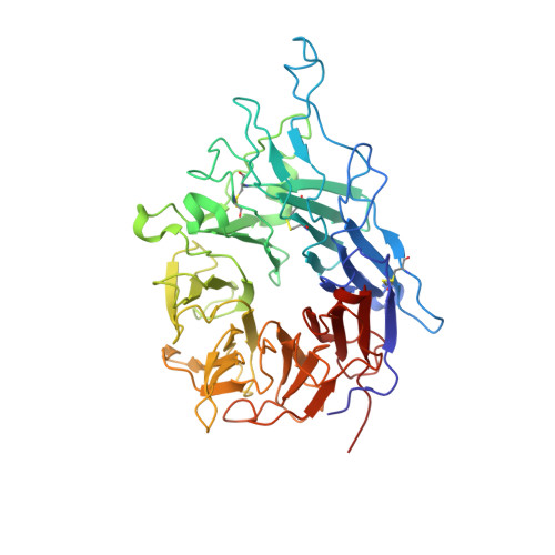
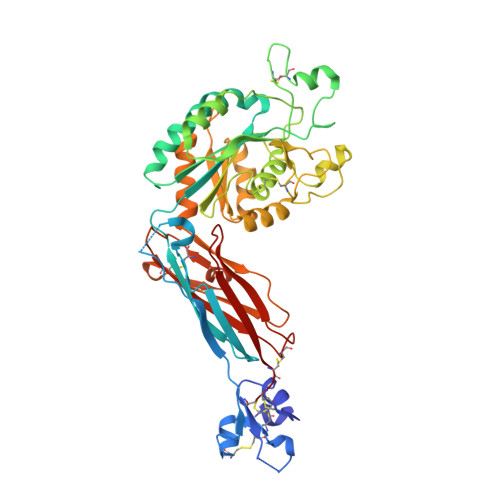
Metal ion and ligand binding of integrin alpha 5 beta 1.

Xia, W.Springer, T.A.

(2014) Proc Natl Acad Sci U S A 111: 17863-17868

  • DOI: https://doi.org/10.1073/pnas.1420645111
  • Primary Citation of Related Structures:  
    4WJK, 4WK0, 4WK2, 4WK4

  • PubMed Abstract: 

    Integrin α5β1 binds to an Arg-Gly-Asp (RGD) motif in its ligand fibronectin. We report high-resolution crystal structures of a four-domain α5β1 headpiece fragment, alone or with RGD peptides soaked into crystals, and RGD peptide affinity measurements. The headpiece crystallizes in a closed conformation essentially identical to that seen previously for α5β1 complexed with a Fab that allosterically inhibits ligand binding by stabilizing the closed conformation. Soaking experiments show that binding of cyclic RGD peptide with 20-fold higher affinity than a linear RGD peptide induces conformational change in the β1-subunit βI domain to a state that is intermediate between closed (low affinity) and open (high affinity). In contrast, binding of a linear RGD peptide induces no shape shifting. However, linear peptide binding induces shape shifting when Ca(2+) is depleted during soaking. Ca(2+) bound to the adjacent to metal ion-dependent adhesion site (ADMIDAS), at the locus of shape shifting, moves and decreases in occupancy, correlating with an increase in affinity for RGD measured when Ca(2+) is depleted. The results directly demonstrate that Ca(2+) binding to the ADMIDAS stabilizes integrins in the low-affinity, closed conformation. Comparisons in affinity between four-domain and six-domain headpiece constructs suggest that flexible integrin leg domains contribute to conformational equilibria. High-resolution views of the hybrid domain interface with the plexin-semaphorin-integrin (PSI) domain in different orientations show a ball-and-socket joint with a hybrid domain Arg side chain that rocks in a PSI domain socket lined with carbonyl oxygens.


  • Organizational Affiliation

    Program in Cellular and Molecular Medicine, Boston Children's Hospital, Boston, MA 02115; and Department of Biological Chemistry and Molecular Pharmacology, Harvard Medical School, Boston, MA 02115.


Macromolecules
Find similar proteins by:  (by identity cutoff)  |  3D Structure
Entity ID: 1
MoleculeChains Sequence LengthOrganismDetailsImage
Integrin alpha-5452Homo sapiensMutation(s): 1 
Gene Names: ITGA5FNRA
UniProt & NIH Common Fund Data Resources
Find proteins for P08648 (Homo sapiens)
Explore P08648 
Go to UniProtKB:  P08648
PHAROS:  P08648
GTEx:  ENSG00000161638 
Entity Groups  
Sequence Clusters30% Identity50% Identity70% Identity90% Identity95% Identity100% Identity
UniProt GroupP08648
Sequence Annotations
Expand
  • Reference Sequence
Find similar proteins by:  (by identity cutoff)  |  3D Structure
Entity ID: 2
MoleculeChains Sequence LengthOrganismDetailsImage
Integrin beta-1445Homo sapiensMutation(s): 0 
Gene Names: ITGB1FNRBMDF2MSK12
UniProt & NIH Common Fund Data Resources
Find proteins for P05556 (Homo sapiens)
Explore P05556 
Go to UniProtKB:  P05556
PHAROS:  P05556
GTEx:  ENSG00000150093 
Entity Groups  
Sequence Clusters30% Identity50% Identity70% Identity90% Identity95% Identity100% Identity
UniProt GroupP05556
Sequence Annotations
Expand
  • Reference Sequence
Oligosaccharides

Help

Entity ID: 3
MoleculeChains Length2D Diagram Glycosylation3D Interactions
alpha-D-mannopyranose-(1-3)-alpha-D-mannopyranose-(1-6)-[alpha-D-mannopyranose-(1-3)]beta-D-mannopyranose-(1-4)-2-acetamido-2-deoxy-beta-D-glucopyranose-(1-4)-2-acetamido-2-deoxy-beta-D-glucopyranose
C
6N-Glycosylation
Glycosylation Resources
GlyTouCan:  G09724ZC
GlyCosmos:  G09724ZC
GlyGen:  G09724ZC
Entity ID: 4
MoleculeChains Length2D Diagram Glycosylation3D Interactions
beta-D-mannopyranose-(1-4)-2-acetamido-2-deoxy-beta-D-glucopyranose-(1-4)-2-acetamido-2-deoxy-beta-D-glucopyranose
D
3N-Glycosylation
Glycosylation Resources
GlyTouCan:  G15407YE
GlyCosmos:  G15407YE
GlyGen:  G15407YE
Entity ID: 5
MoleculeChains Length2D Diagram Glycosylation3D Interactions
alpha-D-mannopyranose-(1-3)-[alpha-D-mannopyranose-(1-6)]alpha-D-mannopyranose-(1-6)-[alpha-D-mannopyranose-(1-3)]beta-D-mannopyranose-(1-4)-2-acetamido-2-deoxy-beta-D-glucopyranose-(1-4)-2-acetamido-2-deoxy-beta-D-glucopyranose
E
7N-Glycosylation
Glycosylation Resources
GlyTouCan:  G55220VL
GlyCosmos:  G55220VL
GlyGen:  G55220VL
Entity ID: 6
MoleculeChains Length2D Diagram Glycosylation3D Interactions
2-acetamido-2-deoxy-beta-D-glucopyranose-(1-4)-2-acetamido-2-deoxy-beta-D-glucopyranose
F, G, H
2N-Glycosylation
Glycosylation Resources
GlyTouCan:  G42666HT
GlyCosmos:  G42666HT
GlyGen:  G42666HT
Entity ID: 7
MoleculeChains Length2D Diagram Glycosylation3D Interactions
alpha-D-mannopyranose-(1-3)-beta-D-mannopyranose-(1-4)-2-acetamido-2-deoxy-beta-D-glucopyranose-(1-4)-2-acetamido-2-deoxy-beta-D-glucopyranose
I
4N-Glycosylation
Glycosylation Resources
GlyTouCan:  G81315DD
GlyCosmos:  G81315DD
GlyGen:  G81315DD
Small Molecules
Ligands 3 Unique
IDChains Name / Formula / InChI Key2D Diagram3D Interactions
NAG
Query on NAG

Download Ideal Coordinates CCD File 
Q [auth B]2-acetamido-2-deoxy-beta-D-glucopyranose
C8 H15 N O6
OVRNDRQMDRJTHS-FMDGEEDCSA-N
CA
Query on CA

Download Ideal Coordinates CCD File 
J [auth A]
K [auth A]
L [auth A]
M [auth A]
O [auth B]
J [auth A],
K [auth A],
L [auth A],
M [auth A],
O [auth B],
P [auth B]
CALCIUM ION
Ca
BHPQYMZQTOCNFJ-UHFFFAOYSA-N
MG
Query on MG

Download Ideal Coordinates CCD File 
N [auth B]MAGNESIUM ION
Mg
JLVVSXFLKOJNIY-UHFFFAOYSA-N
Experimental Data & Validation

Experimental Data

  • Method: X-RAY DIFFRACTION
  • Resolution: 1.85 Å
  • R-Value Free: 0.238 
  • R-Value Work: 0.191 
  • R-Value Observed: 0.192 
  • Space Group: P 21 21 21
Unit Cell:
Length ( Å )Angle ( ˚ )
a = 52.13α = 90
b = 116.75β = 90
c = 159.49γ = 90
Software Package:
Software NamePurpose
XDSdata scaling
PHENIXrefinement
PDB_EXTRACTdata extraction

Structure Validation

View Full Validation Report


Ligand Structure Quality Assessment 


Entry History 

Deposition Data

Revision History  (Full details and data files)

  • Version 1.0: 2014-12-03
    Type: Initial release
  • Version 1.1: 2014-12-17
    Changes: Database references
  • Version 1.2: 2014-12-24
    Changes: Database references
  • Version 2.0: 2017-11-22
    Changes: Atomic model, Database references, Derived calculations, Other, Refinement description, Source and taxonomy, Structure summary
  • Version 3.0: 2020-07-29
    Type: Remediation
    Reason: Carbohydrate remediation
    Changes: Advisory, Atomic model, Data collection, Derived calculations, Refinement description, Structure summary
  • Version 3.1: 2023-12-27
    Changes: Data collection, Database references, Derived calculations, Structure summary