4URA

Crystal structure of human JMJD2A in complex with compound 14a


Experimental Data Snapshot

  • Method: X-RAY DIFFRACTION
  • Resolution: 2.23 Å
  • R-Value Free: 0.259 
  • R-Value Work: 0.207 
  • R-Value Observed: 0.209 

wwPDB Validation   3D Report Full Report


Ligand Structure Quality Assessment 


This is version 1.2 of the entry. See complete history


Literature

Optimisation of a triazolopyridine based histone demethylase inhibitor yields a potent and selective KDM2A (FBXL11) inhibitor.

England, K.S.Tumber, A.Krojer, T.Scozzafava, G.Ng, S.S.Daniel, M.Szykowska, A.Che, K.von Delft, F.Burgess-Brown, N.A.Kawamura, A.Schofield, C.J.Brennan, P.E.

(2014) Medchemcomm 5: 1879-1886

  • DOI: https://doi.org/10.1039/C4MD00291A
  • Primary Citation of Related Structures:  
    4URA

  • PubMed Abstract: 

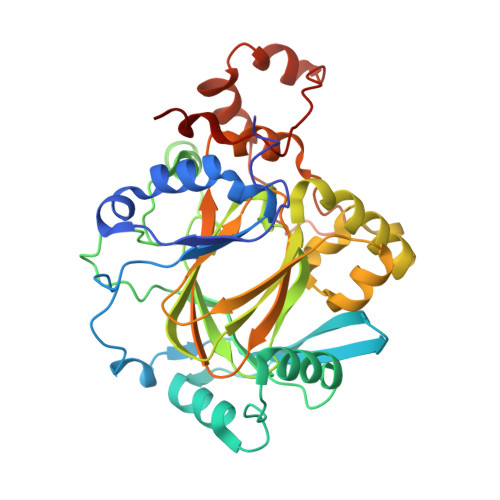
    A potent inhibitor of the JmjC histone lysine demethylase KDM2A (compound 35 , pIC 50 7.2) with excellent selectivity over representatives from other KDM subfamilies has been developed; the discovery that a triazolopyridine compound binds to the active site of JmjC KDMs was followed by optimisation of the triazole substituent for KDM2A inhibition and selectivity.


  • Organizational Affiliation

    Structural Genomics Consortium, University of Oxford, Old Road Campus, Roosevelt Drive, Headington OX3 7DQ, UK.


Macromolecules
Find similar proteins by:  (by identity cutoff)  |  3D Structure
Entity ID: 1
MoleculeChains Sequence LengthOrganismDetailsImage
LYSINE-SPECIFIC DEMETHYLASE 4A
A, B
360Homo sapiensMutation(s): 0 
EC: 1.14.11
UniProt & NIH Common Fund Data Resources
Find proteins for O75164 (Homo sapiens)
Explore O75164 
Go to UniProtKB:  O75164
PHAROS:  O75164
GTEx:  ENSG00000066135 
Entity Groups  
Sequence Clusters30% Identity50% Identity70% Identity90% Identity95% Identity100% Identity
UniProt GroupO75164
Sequence Annotations
Expand
  • Reference Sequence
Small Molecules
Ligands 5 Unique
IDChains Name / Formula / InChI Key2D Diagram3D Interactions
LEL
Query on LEL

Download Ideal Coordinates CCD File 
G [auth A],
K [auth B]
2-(2H-1,2,3-triazol-4-yl)pyridine-4-carboxylic acid
C8 H6 N4 O2
XGBRXPYFGYDEMS-UHFFFAOYSA-N
SO4
Query on SO4

Download Ideal Coordinates CCD File 
D [auth A],
H [auth B]
SULFATE ION
O4 S
QAOWNCQODCNURD-UHFFFAOYSA-L
ZN
Query on ZN

Download Ideal Coordinates CCD File 
F [auth A],
J [auth B]
ZINC ION
Zn
PTFCDOFLOPIGGS-UHFFFAOYSA-N
EDO
Query on EDO

Download Ideal Coordinates CCD File 
C [auth A]1,2-ETHANEDIOL
C2 H6 O2
LYCAIKOWRPUZTN-UHFFFAOYSA-N
NI
Query on NI

Download Ideal Coordinates CCD File 
E [auth A],
I [auth B]
NICKEL (II) ION
Ni
VEQPNABPJHWNSG-UHFFFAOYSA-N
Binding Affinity Annotations 
IDSourceBinding Affinity
LEL Binding MOAD:  4URA IC50: 3.16e-3 (nM) from 1 assay(s)
Experimental Data & Validation

Experimental Data

  • Method: X-RAY DIFFRACTION
  • Resolution: 2.23 Å
  • R-Value Free: 0.259 
  • R-Value Work: 0.207 
  • R-Value Observed: 0.209 
  • Space Group: P 21 21 2
Unit Cell:
Length ( Å )Angle ( ˚ )
a = 101.04α = 90
b = 149.66β = 90
c = 57.86γ = 90
Software Package:
Software NamePurpose
PHENIXrefinement
XDSdata reduction
Aimlessdata scaling

Structure Validation

View Full Validation Report


Ligand Structure Quality Assessment 


Entry History 

Revision History  (Full details and data files)

  • Version 1.0: 2015-06-17
    Type: Initial release
  • Version 1.1: 2017-01-18
    Changes: Database references
  • Version 1.2: 2018-01-24
    Changes: Structure summary