4TVM

Structure of Citrate Synthase from Mycobacterium tuberculosis


Experimental Data Snapshot

  • Method: X-RAY DIFFRACTION
  • Resolution: 2.60 Å
  • R-Value Free: 0.194 
  • R-Value Work: 0.174 
  • R-Value Observed: 0.175 

wwPDB Validation   3D Report Full Report


This is version 1.3 of the entry. See complete history


Literature

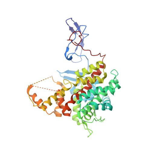
Structures of citrate synthase and malate dehydrogenase of Mycobacterium tuberculosis.

Ferraris, D.M.Spallek, R.Oehlmann, W.Singh, M.Rizzi, M.

(2015) Proteins 83: 389-394

  • DOI: https://doi.org/10.1002/prot.24743
  • Primary Citation of Related Structures:  
    4TVM, 4TVO

  • PubMed Abstract: 

    The tricarboxylic acid (TCA) cycle is a central metabolic pathway of all aerobic organisms and is responsible for the synthesis of many important precursors and molecules. TCA cycle plays a key role in the metabolism of Mycobacterium tuberculosis and is involved in the adaptation process of the bacteria to the host immune response. We present here the first crystal structures of M. tuberculosis malate dehydrogenase and citrate synthase, two consecutive enzymes of the TCA, at 2.6 Å and 1.5 Å resolution, respectively. General analogies and local differences with the previously reported homologous protein structures are described.


  • Organizational Affiliation

    Department of Pharmaceutical Sciences, Università del Piemonte Orientale "A. Avogadro", Largo Donegani 2, 28100, Novara, Italy.


Macromolecules
Find similar proteins by:  (by identity cutoff)  |  3D Structure
Entity ID: 1
MoleculeChains Sequence LengthOrganismDetailsImage
Citrate synthase431Mycobacterium tuberculosis H37RvMutation(s): 0 
Gene Names: RVBD_0896
EC: 2.3.3.1
UniProt
Find proteins for P9WPD5 (Mycobacterium tuberculosis (strain ATCC 25618 / H37Rv))
Explore P9WPD5 
Go to UniProtKB:  P9WPD5
Entity Groups  
Sequence Clusters30% Identity50% Identity70% Identity90% Identity95% Identity100% Identity
UniProt GroupP9WPD5
Sequence Annotations
Expand
  • Reference Sequence
Small Molecules
Ligands 1 Unique
IDChains Name / Formula / InChI Key2D Diagram3D Interactions
OAA
Query on OAA

Download Ideal Coordinates CCD File 
B [auth A]OXALOACETATE ION
C4 H3 O5
KHPXUQMNIQBQEV-UHFFFAOYSA-M
Experimental Data & Validation

Experimental Data

  • Method: X-RAY DIFFRACTION
  • Resolution: 2.60 Å
  • R-Value Free: 0.194 
  • R-Value Work: 0.174 
  • R-Value Observed: 0.175 
  • Space Group: H 3 2
Unit Cell:
Length ( Å )Angle ( ˚ )
a = 148.767α = 90
b = 148.767β = 90
c = 182.797γ = 120
Software Package:
Software NamePurpose
REFMACrefinement

Structure Validation

View Full Validation Report


Entry History & Funding Information

Deposition Data


Funding OrganizationLocationGrant Number
European Union FP7 ProgramItalySysteMTb HEALTH-F4-2010-241587

Revision History  (Full details and data files)

  • Version 1.0: 2015-01-14
    Type: Initial release
  • Version 1.1: 2015-01-28
    Changes: Database references
  • Version 1.2: 2019-10-16
    Changes: Data collection
  • Version 1.3: 2023-12-20
    Changes: Data collection, Database references, Refinement description