4REK

Crystal structure and charge density studies of cholesterol oxidase from Brevibacterium sterolicum at 0.74 ultra-high resolution


Experimental Data Snapshot

  • Method: X-RAY DIFFRACTION
  • Resolution: 0.74 Å
  • R-Value Free: 0.123 
  • R-Value Work: 0.113 
  • R-Value Observed: 0.113 

wwPDB Validation   3D Report Full Report


Ligand Structure Quality Assessment 


This is version 1.3 of the entry. See complete history


Literature

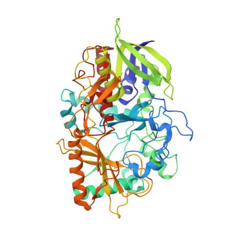
Cholesterol oxidase: ultrahigh-resolution crystal structure and multipolar atom model-based analysis.

Zarychta, B.Lyubimov, A.Ahmed, M.Munshi, P.Guillot, B.Vrielink, A.Jelsch, C.

(2015) Acta Crystallogr D Biol Crystallogr 71: 954-968

  • DOI: https://doi.org/10.1107/S1399004715002382
  • Primary Citation of Related Structures:  
    4REK

  • PubMed Abstract: 

    Examination of protein structure at the subatomic level is required to improve the understanding of enzymatic function. For this purpose, X-ray diffraction data have been collected at 100 K from cholesterol oxidase crystals using synchrotron radiation to an optical resolution of 0.94 Å. After refinement using the spherical atom model, nonmodelled bonding peaks were detected in the Fourier residual electron density on some of the individual bonds. Well defined bond density was observed in the peptide plane after averaging maps on the residues with the lowest thermal motion. The multipolar electron density of the protein-cofactor complex was modelled by transfer of the ELMAM2 charge-density database, and the topology of the intermolecular interactions between the protein and the flavin adenine dinucleotide (FAD) cofactor was subsequently investigated. Taking advantage of the high resolution of the structure, the stereochemistry of main-chain bond lengths and of C=O···H-N hydrogen bonds was analyzed with respect to the different secondary-structure elements.


  • Organizational Affiliation

    Laboratoire de Cristallographie, Résonance Magnétique et Modélisations (CRM2), CNRS, UMR 7036, Institut Jean Barriol, Faculté des Sciences et Technologies, Université de Lorraine, BP 70239, 54506 Vandoeuvre-lès-Nancy CEDEX, France.


Macromolecules
Find similar proteins by:  (by identity cutoff)  |  3D Structure
Entity ID: 1
MoleculeChains Sequence LengthOrganismDetailsImage
Cholesterol oxidase499Streptomyces sp. SA-COOMutation(s): 0 
Gene Names: choA
EC: 1.1.3.6 (PDB Primary Data), 5.3.3.1 (PDB Primary Data)
UniProt
Find proteins for P12676 (Streptomyces sp. (strain SA-COO))
Explore P12676 
Go to UniProtKB:  P12676
Entity Groups  
Sequence Clusters30% Identity50% Identity70% Identity90% Identity95% Identity100% Identity
UniProt GroupP12676
Sequence Annotations
Expand
  • Reference Sequence
Small Molecules
Ligands 2 Unique
IDChains Name / Formula / InChI Key2D Diagram3D Interactions
FAD
Query on FAD

Download Ideal Coordinates CCD File 
B [auth A]FLAVIN-ADENINE DINUCLEOTIDE
C27 H33 N9 O15 P2
VWWQXMAJTJZDQX-UYBVJOGSSA-N
PO4
Query on PO4

Download Ideal Coordinates CCD File 
C [auth A]PHOSPHATE ION
O4 P
NBIIXXVUZAFLBC-UHFFFAOYSA-K
Experimental Data & Validation

Experimental Data

  • Method: X-RAY DIFFRACTION
  • Resolution: 0.74 Å
  • R-Value Free: 0.123 
  • R-Value Work: 0.113 
  • R-Value Observed: 0.113 
  • Space Group: P 1 21 1
Unit Cell:
Length ( Å )Angle ( ˚ )
a = 51.24α = 90
b = 72.92β = 105.13
c = 63.01γ = 90
Software Package:
Software NamePurpose
Blu-Icedata collection
PHENIXrefinement
d*TREKdata reduction
CCP4data scaling

Structure Validation

View Full Validation Report


Ligand Structure Quality Assessment 


Entry History 

Deposition Data

Revision History  (Full details and data files)

  • Version 1.0: 2015-04-15
    Type: Initial release
  • Version 1.1: 2015-06-03
    Changes: Database references
  • Version 1.2: 2017-11-22
    Changes: Refinement description
  • Version 1.3: 2023-09-20
    Changes: Data collection, Database references, Derived calculations, Refinement description