4O7D

Crystal structure of human glutaminase in complex DON


Experimental Data Snapshot

  • Method: X-RAY DIFFRACTION
  • Resolution: 2.30 Å
  • R-Value Free: 0.211 
  • R-Value Work: 0.193 
  • R-Value Observed: 0.194 

wwPDB Validation   3D Report Full Report


This is version 1.0 of the entry. See complete history


Literature

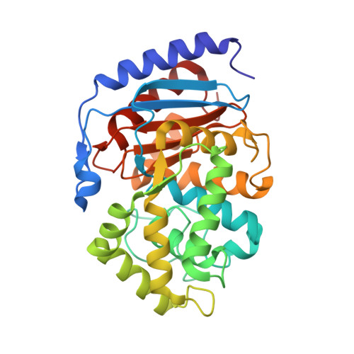
Structural basis for the active site inhibition mechanism of human kidney-type glutaminase (KGA)

Thangavelu, K.Chong, Q.Y.Low, B.C.Sivaraman, J.

(2014) Sci Rep 4: 3827-3827

  • DOI: https://doi.org/10.1038/srep03827
  • Primary Citation of Related Structures:  
    4O7D

  • PubMed Abstract: 

    Glutaminase is a metabolic enzyme responsible for glutaminolysis, a process harnessed by cancer cells to feed their accelerated growth and proliferation. Among the glutaminase isoforms, human kidney-type glutaminase (KGA) is often upregulated in cancer and is thus touted as an attractive drug target. Here we report the active site inhibition mechanism of KGA through the crystal structure of the catalytic domain of KGA (cKGA) in complex with 6-diazo-5-oxo-L-norleucine (DON), a substrate analogue of glutamine. DON covalently binds with the active site Ser286 and interacts with residues such as Tyr249, Asn335, Glu381, Asn388, Tyr414, Tyr466 and Val484. The nucleophilic attack of Ser286 sidechain on DON releases the diazo group (N2) from the inhibitor and results in the formation of an enzyme-inhibitor complex. Mutational studies confirmed the key role of these residues in the activity of KGA. This study will be important in the development of KGA active site inhibitors for therapeutic interventions.


  • Organizational Affiliation

    Department of Biological Sciences, 14 Science Drive 4, National University of Singapore, Singapore 117543.


Macromolecules
Find similar proteins by:  (by identity cutoff)  |  3D Structure
Entity ID: 1
MoleculeChains Sequence LengthOrganismDetailsImage
Glutaminase kidney isoform, mitochondrial313Homo sapiensMutation(s): 0 
Gene Names: GLSGLS1KIAA0838
EC: 3.5.1.2
UniProt & NIH Common Fund Data Resources
Find proteins for O94925 (Homo sapiens)
Explore O94925 
Go to UniProtKB:  O94925
PHAROS:  O94925
GTEx:  ENSG00000115419 
Entity Groups  
Sequence Clusters30% Identity50% Identity70% Identity90% Identity95% Identity100% Identity
UniProt GroupO94925
Sequence Annotations
Expand
  • Reference Sequence
Small Molecules
Ligands 1 Unique
IDChains Name / Formula / InChI Key2D Diagram3D Interactions
ONL
Query on ONL

Download Ideal Coordinates CCD File 
B [auth A]5-OXO-L-NORLEUCINE
C6 H11 N O3
KSIJECNNZVKMJG-YFKPBYRVSA-N
Experimental Data & Validation

Experimental Data

  • Method: X-RAY DIFFRACTION
  • Resolution: 2.30 Å
  • R-Value Free: 0.211 
  • R-Value Work: 0.193 
  • R-Value Observed: 0.194 
  • Space Group: I 41 2 2
Unit Cell:
Length ( Å )Angle ( ˚ )
a = 139.278α = 90
b = 139.278β = 90
c = 155.502γ = 90
Software Package:
Software NamePurpose
ADSCdata collection
MOLREPphasing
PHENIXrefinement
HKL-2000data reduction
HKL-2000data scaling

Structure Validation

View Full Validation Report


Entry History 

Deposition Data

Revision History  (Full details and data files)

  • Version 1.0: 2014-04-16
    Type: Initial release