4NE5

Human MHF1-MHF2 complex


Experimental Data Snapshot

  • Method: X-RAY DIFFRACTION
  • Resolution: 2.50 Å
  • R-Value Free: 0.264 
  • R-Value Work: 0.230 
  • R-Value Observed: 0.231 

wwPDB Validation   3D Report Full Report


This is version 1.1 of the entry. See complete history


Literature

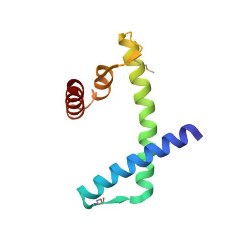
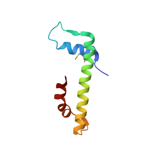
The MHF complex senses branched DNA by binding a pair of crossover DNA duplexes.

Zhao, Q.Saro, D.Sachpatzidis, A.Singh, T.R.Schlingman, D.Zheng, X.F.Mack, A.Tsai, M.S.Mochrie, S.Regan, L.Meetei, A.R.Sung, P.Xiong, Y.

(2014) Nat Commun 5: 2987-2987

  • DOI: https://doi.org/10.1038/ncomms3987
  • Primary Citation of Related Structures:  
    4NDY, 4NE1, 4NE3, 4NE5, 4NE6

  • PubMed Abstract: 

    The conserved MHF1-MHF2 (MHF) complex functions in the activation of the Fanconi anaemia pathway of the DNA damage response, in regulating homologous recombination, and in DNA replication fork maintenance. MHF facilitates the processing of multiple types of branched DNAs by the DNA translocase FANCM. Here we report the crystal structure of a human MHF-DNA complex that reveals the DNA-binding mode of MHF. The structure suggests that MHF prefers branched DNA over double-stranded DNA because it engages two duplex arms. Biochemical analyses verify that MHF preferentially engages DNA forks or various four-way junctions independent of the junction-site structure. Furthermore, genetic experiments provide evidence that the observed DNA-binding interface of MHF is important for cellular resistance to DNA damage. These results offer insights into how the MHF complex recognizes branched DNA and stimulates FANCM activity at such a structure to promote genome maintenance.


  • Organizational Affiliation

    1] Department of Molecular Biophysics and Biochemistry, Yale University School of Medicine, New Haven, Connecticut 06520, USA [2].


Macromolecules
Find similar proteins by:  (by identity cutoff)  |  3D Structure
Entity ID: 1
MoleculeChains Sequence LengthOrganismDetailsImage
Centromere protein S
A, C, E, G
93Homo sapiensMutation(s): 0 
Gene Names: APITD1CENPSFAAP16MHF1
UniProt & NIH Common Fund Data Resources
Find proteins for Q8N2Z9 (Homo sapiens)
Explore Q8N2Z9 
Go to UniProtKB:  Q8N2Z9
PHAROS:  Q8N2Z9
GTEx:  ENSG00000175279 
Entity Groups  
Sequence Clusters30% Identity50% Identity70% Identity90% Identity95% Identity100% Identity
UniProt GroupQ8N2Z9
Sequence Annotations
Expand
  • Reference Sequence
Find similar proteins by:  (by identity cutoff)  |  3D Structure
Entity ID: 2
MoleculeChains Sequence LengthOrganismDetailsImage
Centromere protein X
B, D, F, H
74Homo sapiensMutation(s): 0 
Gene Names: STRA13CENPXFAAP10MHF2
UniProt & NIH Common Fund Data Resources
Find proteins for A8MT69 (Homo sapiens)
Explore A8MT69 
Go to UniProtKB:  A8MT69
PHAROS:  A8MT69
GTEx:  ENSG00000169689 
Entity Groups  
Sequence Clusters30% Identity50% Identity70% Identity90% Identity95% Identity100% Identity
UniProt GroupA8MT69
Sequence Annotations
Expand
  • Reference Sequence
Small Molecules
Modified Residues  1 Unique
IDChains TypeFormula2D DiagramParent
MSE
Query on MSE
A, C, E, G
L-PEPTIDE LINKINGC5 H11 N O2 SeMET
Experimental Data & Validation

Experimental Data

  • Method: X-RAY DIFFRACTION
  • Resolution: 2.50 Å
  • R-Value Free: 0.264 
  • R-Value Work: 0.230 
  • R-Value Observed: 0.231 
  • Space Group: P 1 21 1
Unit Cell:
Length ( Å )Angle ( ˚ )
a = 41.076α = 90
b = 128.76β = 100.96
c = 88.836γ = 90
Software Package:
Software NamePurpose
PHENIXrefinement
PDB_EXTRACTdata extraction
ADSCdata collection
HKL-2000data reduction
HKL-2000data scaling
PHASERphasing

Structure Validation

View Full Validation Report


Entry History 

Deposition Data

Revision History  (Full details and data files)

  • Version 1.0: 2013-12-25
    Type: Initial release
  • Version 1.1: 2014-02-12
    Changes: Database references