4M17

Crystal Structure of Surfactant Protein-D D325A/R343V mutant


Experimental Data Snapshot

  • Method: X-RAY DIFFRACTION
  • Resolution: 2.10 Å
  • R-Value Free: 0.188 
  • R-Value Work: 0.163 
  • R-Value Observed: 0.170 

wwPDB Validation   3D Report Full Report


This is version 1.2 of the entry. See complete history


Literature

Molecular mechanisms of inhibition of influenza by surfactant protein d revealed by large-scale molecular dynamics simulation.

Goh, B.C.Rynkiewicz, M.J.Cafarella, T.R.White, M.R.Hartshorn, K.L.Allen, K.Crouch, E.C.Calin, O.Seeberger, P.H.Schulten, K.Seaton, B.A.

(2013) Biochemistry 52: 8527-8538

  • DOI: https://doi.org/10.1021/bi4010683
  • Primary Citation of Related Structures:  
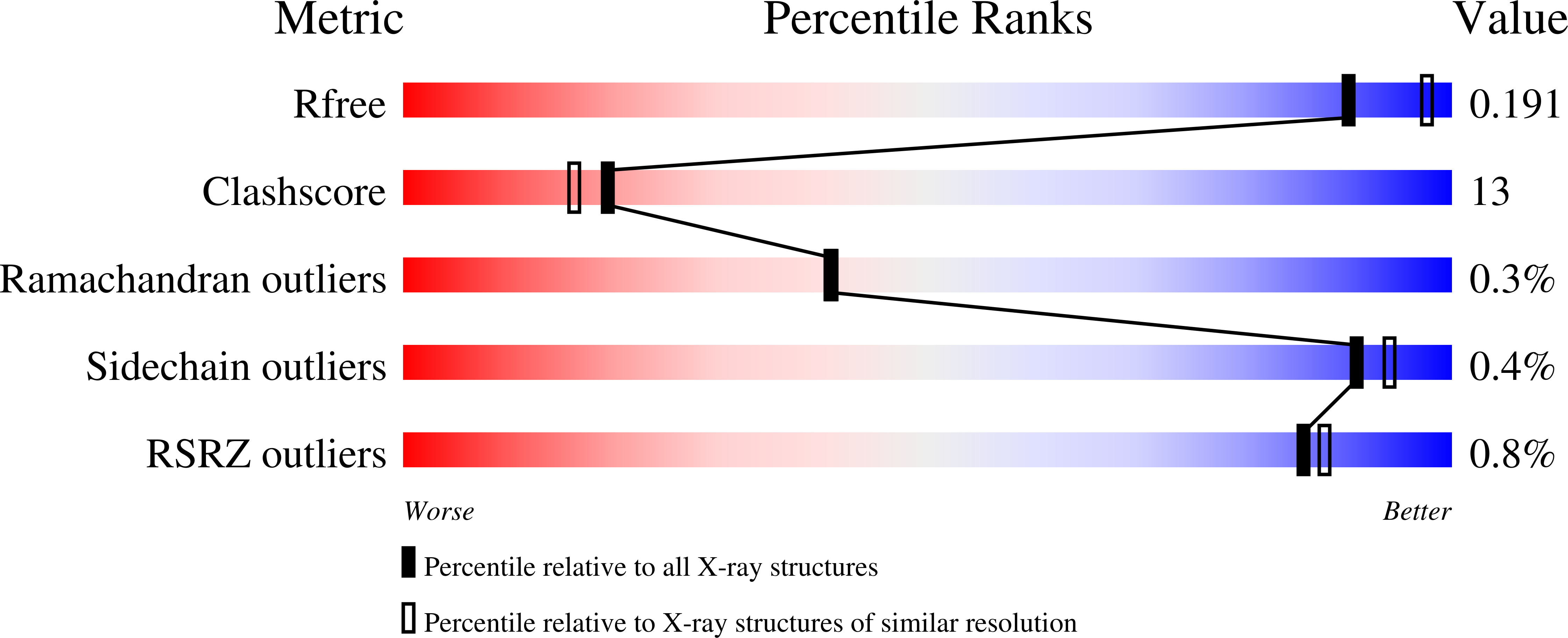
    4M17, 4M18

  • PubMed Abstract: 

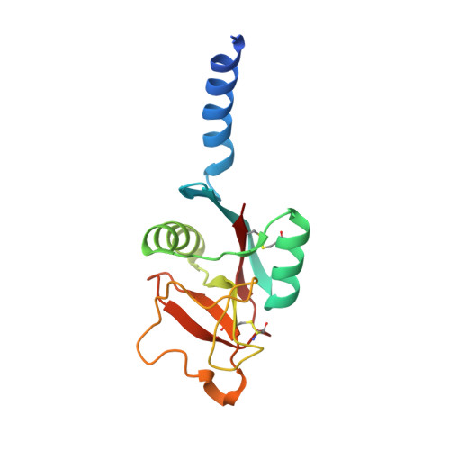
    Surfactant protein D (SP-D), a mammalian C-type lectin, is the primary innate inhibitor of influenza A virus (IAV) in the lung. Interactions of SP-D with highly branched viral N-linked glycans on hemagglutinin (HA), an abundant IAV envelope protein and critical virulence factor, promote viral aggregation and neutralization through as yet unknown molecular mechanisms. Two truncated human SP-D forms, wild-type (WT) and double mutant D325A+R343V, representing neck and carbohydrate recognition domains are compared in this study. Whereas both WT and D325A+R343V bind to isolated glycosylated HA, WT does not inhibit IAV in neutralization assays; in contrast, D325A+R343V neutralization compares well with that of full-length native SP-D. To elucidate the mechanism for these biochemical observations, we have determined crystal structures of D325A+R343V in the presence and absence of a viral nonamannoside (Man9). On the basis of the D325A+R343V-Man9 structure and other crystallographic data, models of complexes between HA and WT or D325A+R343V were produced and subjected to molecular dynamics. Simulations reveal that whereas WT and D325A+R343V both block the sialic acid receptor site of HA, the D325A+R343V complex is more stable, with stronger binding caused by additional hydrogen bonds and hydrophobic interactions with HA residues. Furthermore, the blocking mechanism of HA differs for WT and D325A+R343V because of alternate glycan binding modes. The combined results suggest a mechanism through which the mode of SP-D-HA interaction could significantly influence viral aggregation and neutralization. These studies provide the first atomic-level molecular view of an innate host defense lectin inhibiting its viral glycoprotein target.


  • Organizational Affiliation

    Beckman Institute and Department of Physics, University of Illinois at Urbana-Champaign , Urbana, Illinois 61801, United States.


Macromolecules
Find similar proteins by:  (by identity cutoff)  |  3D Structure
Entity ID: 1
MoleculeChains Sequence LengthOrganismDetailsImage
Pulmonary surfactant-associated protein D
A, B, C, D, E
A, B, C, D, E, F, G, H, I, J, K, L
147Homo sapiensMutation(s): 2 
Gene Names: COLEC7PSPDSFTP4SFTPD
UniProt & NIH Common Fund Data Resources
Find proteins for P35247 (Homo sapiens)
Explore P35247 
Go to UniProtKB:  P35247
PHAROS:  P35247
GTEx:  ENSG00000133661 
Entity Groups  
Sequence Clusters30% Identity50% Identity70% Identity90% Identity95% Identity100% Identity
UniProt GroupP35247
Sequence Annotations
Expand
  • Reference Sequence
Small Molecules
Ligands 1 Unique
IDChains Name / Formula / InChI Key2D Diagram3D Interactions
CA
Query on CA

Download Ideal Coordinates CCD File 
AA [auth E]
BA [auth F]
CA [auth F]
DA [auth F]
EA [auth G]
AA [auth E],
BA [auth F],
CA [auth F],
DA [auth F],
EA [auth G],
FA [auth G],
GA [auth G],
HA [auth H],
IA [auth H],
JA [auth H],
KA [auth I],
LA [auth I],
M [auth A],
MA [auth I],
N [auth A],
NA [auth J],
O [auth A],
OA [auth J],
P [auth B],
PA [auth J],
Q [auth B],
QA [auth K],
R [auth B],
RA [auth K],
S [auth C],
SA [auth K],
T [auth C],
TA [auth L],
U [auth C],
UA [auth L],
V [auth D],
VA [auth L],
W [auth D],
X [auth D],
Y [auth E],
Z [auth E]
CALCIUM ION
Ca
BHPQYMZQTOCNFJ-UHFFFAOYSA-N
Experimental Data & Validation

Experimental Data

  • Method: X-RAY DIFFRACTION
  • Resolution: 2.10 Å
  • R-Value Free: 0.188 
  • R-Value Work: 0.163 
  • R-Value Observed: 0.170 
  • Space Group: P 21 21 21
Unit Cell:
Length ( Å )Angle ( ˚ )
a = 107.524α = 90
b = 160.108β = 90
c = 160.073γ = 90
Software Package:
Software NamePurpose
DENZOdata reduction
SCALEPACKdata scaling
PHASERphasing
PHENIXrefinement
PDB_EXTRACTdata extraction

Structure Validation

View Full Validation Report


Entry History 

Revision History  (Full details and data files)

  • Version 1.0: 2013-12-04
    Type: Initial release
  • Version 1.1: 2013-12-11
    Changes: Database references
  • Version 1.2: 2023-09-20
    Changes: Data collection, Database references, Derived calculations, Refinement description