4KUO

A superfast recovering full-length LOV protein from the marine phototrophic bacterium Dinoroseobacter shibae (Photoexcited state)


Experimental Data Snapshot

  • Method: X-RAY DIFFRACTION
  • Resolution: 2.00 Å
  • R-Value Free: 0.182 
  • R-Value Work: 0.163 
  • R-Value Observed: 0.164 

wwPDB Validation   3D Report Full Report


Ligand Structure Quality Assessment 


This is version 1.4 of the entry. See complete history


Literature

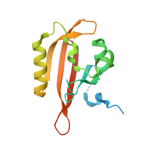
Structure and function of a short LOV protein from the marine phototrophic bacterium Dinoroseobacter shibae.

Endres, S.Granzin, J.Circolone, F.Stadler, A.Krauss, U.Drepper, T.Svensson, V.Knieps-Grunhagen, E.Wirtz, A.Cousin, A.Tielen, P.Willbold, D.Jaeger, K.E.Batra-Safferling, R.

(2015) BMC Microbiol 15: 30-30

  • DOI: https://doi.org/10.1186/s12866-015-0365-0
  • Primary Citation of Related Structures:  
    4KUK, 4KUO

  • PubMed Abstract: 

    Light, oxygen, voltage (LOV) domains are widely distributed in plants, algae, fungi, bacteria, and represent the photo-responsive domains of various blue-light photoreceptor proteins. Their photocycle involves the blue-light triggered adduct formation between the C(4a) atom of a non-covalently bound flavin chromophore and the sulfur atom of a conserved cysteine in the LOV sensor domain. LOV proteins show considerable variation in the structure of N- and C-terminal elements which flank the LOV core domain, as well as in the lifetime of the adduct state.


  • Organizational Affiliation

    Institute of Molecular Enzyme Technology, Heinrich-Heine-Universität Düsseldorf, Forschungszentrum Jülich, D-52425, Jülich, Germany. s.endres@fz-juelich.de.


Macromolecules
Find similar proteins by:  (by identity cutoff)  |  3D Structure
Entity ID: 1
MoleculeChains Sequence LengthOrganismDetailsImage
blue-light photoreceptor146Dinoroseobacter shibae DFL 12 = DSM 16493Mutation(s): 0 
Gene Names: Dshi_2006
UniProt
Find proteins for A8LP63 (Dinoroseobacter shibae (strain DSM 16493 / NCIMB 14021 / DFL 12))
Explore A8LP63 
Go to UniProtKB:  A8LP63
Entity Groups  
Sequence Clusters30% Identity50% Identity70% Identity90% Identity95% Identity100% Identity
UniProt GroupA8LP63
Sequence Annotations
Expand
  • Reference Sequence
Small Molecules
Ligands 1 Unique
IDChains Name / Formula / InChI Key2D Diagram3D Interactions
RBF
Query on RBF

Download Ideal Coordinates CCD File 
B [auth A]RIBOFLAVIN
C17 H20 N4 O6
AUNGANRZJHBGPY-SCRDCRAPSA-N
Experimental Data & Validation

Experimental Data

  • Method: X-RAY DIFFRACTION
  • Resolution: 2.00 Å
  • R-Value Free: 0.182 
  • R-Value Work: 0.163 
  • R-Value Observed: 0.164 
  • Space Group: C 1 2 1
Unit Cell:
Length ( Å )Angle ( ˚ )
a = 90.78α = 90
b = 30.936β = 113.09
c = 49.538γ = 90
Software Package:
Software NamePurpose
ADSCdata collection
PHASERphasing
PHENIXrefinement
MOSFLMdata reduction
SCALAdata scaling

Structure Validation

View Full Validation Report


Ligand Structure Quality Assessment 


Entry History 

Revision History  (Full details and data files)

  • Version 1.0: 2014-11-26
    Type: Initial release
  • Version 1.1: 2015-03-04
    Changes: Database references
  • Version 1.2: 2015-04-01
    Changes: Database references
  • Version 1.3: 2015-04-29
    Changes: Database references
  • Version 1.4: 2023-09-20
    Changes: Data collection, Database references, Derived calculations, Refinement description