4JQH

Crystal structure of a new sGC activator (analogue of BAY 58-2667) bound to nostoc H-NOX domain


Experimental Data Snapshot

  • Method: X-RAY DIFFRACTION
  • Resolution: 2.30 Å
  • R-Value Free: 0.190 
  • R-Value Work: 0.129 
  • R-Value Observed: 0.132 

wwPDB Validation   3D Report Full Report


Ligand Structure Quality Assessment 


This is version 1.3 of the entry. See complete history


Literature

Insights into soluble guanylyl cyclase activation derived from improved heme-mimetics.

von Wantoch Rekowski, M.Kumar, V.Zhou, Z.Moschner, J.Marazioti, A.Bantzi, M.Spyroulias, G.A.van den Akker, F.Giannis, A.Papapetropoulos, A.

(2013) J Med Chem 56: 8948-8952

  • DOI: https://doi.org/10.1021/jm400539d
  • Primary Citation of Related Structures:  
    4JQH

  • PubMed Abstract: 

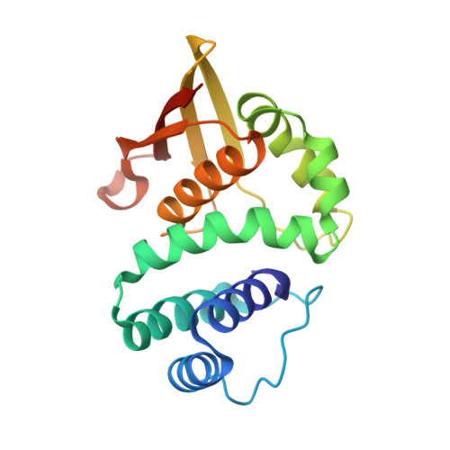
    Recently, the structure of BAY 58-2667 bound to the Nostoc sp. H-NOX domain was published. On the basis of this structural information, we designed BAY 58-2667 derivatives and tested their effects on soluble guanylyl cyclase (sGC) activity. Derivative 20 activated sGC 4.8-fold more than BAY 58-2667. Co-crystallization of 20 with the Ns H-NOX domain revealed that the increased conformational distortion at the C-terminal region of αF helix containing 110-114 residues contributes to the higher activation triggered by 20.


  • Organizational Affiliation

    Institut für Organische Chemie, Universität Leipzig, Leipzig, Germany.


Macromolecules
Find similar proteins by:  (by identity cutoff)  |  3D Structure
Entity ID: 1
MoleculeChains Sequence LengthOrganismDetailsImage
Alr2278 protein
A, B
189Nostoc sp. PCC 7120 = FACHB-418Mutation(s): 1 
Gene Names: alr2278
EC: 4.6.1.2
UniProt
Find proteins for Q8YUQ7 (Nostoc sp. (strain PCC 7120 / SAG 25.82 / UTEX 2576))
Explore Q8YUQ7 
Go to UniProtKB:  Q8YUQ7
Entity Groups  
Sequence Clusters30% Identity50% Identity70% Identity90% Identity95% Identity100% Identity
UniProt GroupQ8YUQ7
Sequence Annotations
Expand
  • Reference Sequence
Experimental Data & Validation

Experimental Data

  • Method: X-RAY DIFFRACTION
  • Resolution: 2.30 Å
  • R-Value Free: 0.190 
  • R-Value Work: 0.129 
  • R-Value Observed: 0.132 
  • Space Group: P 21 3
Unit Cell:
Length ( Å )Angle ( ˚ )
a = 122.191α = 90
b = 122.191β = 90
c = 122.191γ = 90
Software Package:
Software NamePurpose
HKL-2000data collection
MOLREPphasing
REFMACrefinement
HKL-2000data reduction
HKL-2000data scaling

Structure Validation

View Full Validation Report


Ligand Structure Quality Assessment 


Entry History 

Deposition Data

Revision History  (Full details and data files)

  • Version 1.0: 2013-11-20
    Type: Initial release
  • Version 1.1: 2014-01-15
    Changes: Database references
  • Version 1.2: 2015-04-29
    Changes: Non-polymer description
  • Version 1.3: 2024-02-28
    Changes: Data collection, Database references, Derived calculations