4HHV

Crystal structure of ceramide transfer protein pleckstrin homology domain


Experimental Data Snapshot

  • Method: X-RAY DIFFRACTION
  • Resolution: 1.75 Å
  • R-Value Free: 0.204 
  • R-Value Work: 0.179 
  • R-Value Observed: 0.180 

wwPDB Validation   3D Report Full Report


This is version 1.2 of the entry. See complete history


Literature

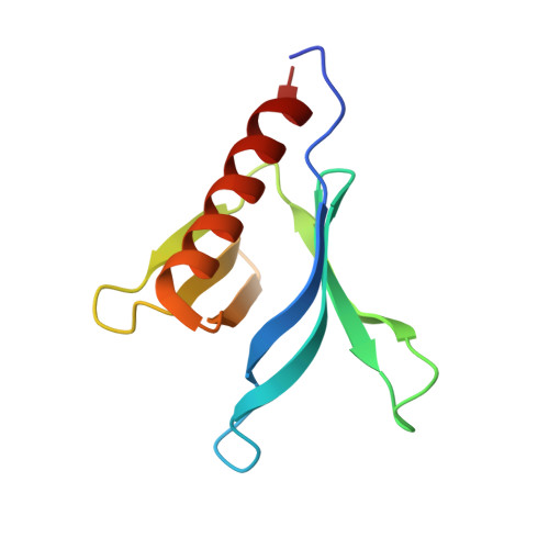
Crystal Structure of the Pleckstrin Homology Domain from the Ceramide Transfer Protein: Implications for Conformational Change upon Ligand Binding.

Prashek, J.Truong, T.Yao, X.

(2013) PLoS One 8: e79590-e79590

  • DOI: https://doi.org/10.1371/journal.pone.0079590
  • Primary Citation of Related Structures:  
    4HHV

  • PubMed Abstract: 

    Ceramide transfer protein (CERT) is responsible for the nonvesicular trafficking of ceramide from the endoplasmic reticulum (ER) to the trans Golgi network where it is converted to sphingomyelin (SM). The N-terminal pleckstrin homology (PH) domain is required for Golgi targeting of CERT by recognizing the phosphatidylinositol 4-phosphate (PtdIns(4)P) enriched in the Golgi membrane. We report a crystal structure of the CERT PH domain. This structure contains a sulfate that is hydrogen bonded with residues in the canonical ligand-binding pocket of PH domains. Our nuclear magnetic resonance (NMR) chemical shift perturbation (CSP) analyses show sulfate association with CERT PH protein resembles that of PtdIns(4)P, suggesting that the sulfate bound structure likely mimics the holo form of CERT PH protein. Comparison of the sulfate bound structure with the apo form solution structure shows structural rearrangements likely occur upon ligand binding, suggesting conformational flexibility in the ligand-binding pocket. This structural flexibility likely explains CERT PH domain's low affinity for PtdIns(4)P, a property that is distinct from many other PH domains that bind to their phosphoinositide ligands tightly. This unique structural feature of CERT PH domain is probably tailored towards the transfer activity of CERT protein where it needs to shuttle between ER and Golgi and therefore requires short resident time on ER and Golgi membranes.


  • Organizational Affiliation

    Division of Molecular Biology and Biochemistry, School of Biological Sciences, University of Missouri Kansas City, Kansas City, Missouri, United States of America.


Macromolecules
Find similar proteins by:  (by identity cutoff)  |  3D Structure
Entity ID: 1
MoleculeChains Sequence LengthOrganismDetailsImage
Collagen type IV alpha-3-binding protein
A, B
105Homo sapiensMutation(s): 0 
Gene Names: CERTCOL4A3BPSTARD11
UniProt & NIH Common Fund Data Resources
Find proteins for Q9Y5P4 (Homo sapiens)
Explore Q9Y5P4 
Go to UniProtKB:  Q9Y5P4
PHAROS:  Q9Y5P4
GTEx:  ENSG00000113163 
Entity Groups  
Sequence Clusters30% Identity50% Identity70% Identity90% Identity95% Identity100% Identity
UniProt GroupQ9Y5P4
Sequence Annotations
Expand
  • Reference Sequence
Small Molecules
Ligands 4 Unique
IDChains Name / Formula / InChI Key2D Diagram3D Interactions
SO4
Query on SO4

Download Ideal Coordinates CCD File 
C [auth A],
G [auth B]
SULFATE ION
O4 S
QAOWNCQODCNURD-UHFFFAOYSA-L
GOL
Query on GOL

Download Ideal Coordinates CCD File 
D [auth A],
H [auth B],
I [auth B]
GLYCEROL
C3 H8 O3
PEDCQBHIVMGVHV-UHFFFAOYSA-N
ACT
Query on ACT

Download Ideal Coordinates CCD File 
K [auth B]ACETATE ION
C2 H3 O2
QTBSBXVTEAMEQO-UHFFFAOYSA-M
CL
Query on CL

Download Ideal Coordinates CCD File 
E [auth A],
F [auth A],
J [auth B]
CHLORIDE ION
Cl
VEXZGXHMUGYJMC-UHFFFAOYSA-M
Experimental Data & Validation

Experimental Data

  • Method: X-RAY DIFFRACTION
  • Resolution: 1.75 Å
  • R-Value Free: 0.204 
  • R-Value Work: 0.179 
  • R-Value Observed: 0.180 
  • Space Group: P 21 21 21
Unit Cell:
Length ( Å )Angle ( ˚ )
a = 48.131α = 90
b = 54.979β = 90
c = 98.988γ = 90
Software Package:
Software NamePurpose
SCALEPACKdata scaling
PHASERphasing
REFMACrefinement
PDB_EXTRACTdata extraction
HKL-2000data collection
HKL-2000data reduction

Structure Validation

View Full Validation Report


Entry History 

Deposition Data

Revision History  (Full details and data files)

  • Version 1.0: 2013-11-27
    Type: Initial release
  • Version 1.1: 2013-12-25
    Changes: Database references
  • Version 1.2: 2024-02-28
    Changes: Data collection, Database references, Derived calculations, Refinement description