4EZM

Crystal structure of the human IgE-Fc(epsilon)3-4 bound to its B cell receptor derCD23


Experimental Data Snapshot

  • Method: X-RAY DIFFRACTION
  • Resolution: 3.10 Å
  • R-Value Free: 0.283 
  • R-Value Work: 0.264 
  • R-Value Observed: 0.265 

wwPDB Validation   3D Report Full Report


Ligand Structure Quality Assessment 


This is version 2.1 of the entry. See complete history


Literature

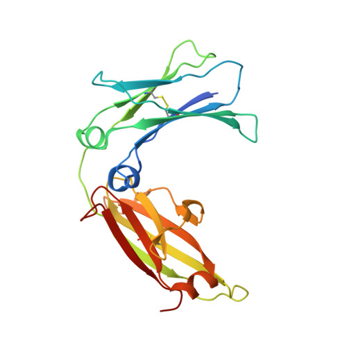
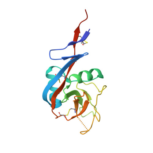
Crystal structure of IgE bound to its B-cell receptor CD23 reveals a mechanism of reciprocal allosteric inhibition with high affinity receptor Fc{varepsilon}RI.

Dhaliwal, B.Yuan, D.Pang, M.O.Henry, A.J.Cain, K.Oxbrow, A.Fabiane, S.M.Beavil, A.J.McDonnell, J.M.Gould, H.J.Sutton, B.J.

(2012) Proc Natl Acad Sci U S A 109: 12686-12691

  • DOI: https://doi.org/10.1073/pnas.1207278109
  • Primary Citation of Related Structures:  
    4EZM

  • PubMed Abstract: 

    The role of IgE in allergic disease mechanisms is performed principally through its interactions with two receptors, FcεRI on mast cells and basophils, and CD23 (FcεRII) on B cells. The former mediates allergic hypersensitivity, the latter regulates IgE levels, and both receptors, also expressed on antigen-presenting cells, contribute to allergen uptake and presentation to the immune system. We have solved the crystal structure of the soluble lectin-like "head" domain of CD23 (derCD23) bound to a subfragment of IgE-Fc consisting of the dimer of Cε3 and Cε4 domains (Fcε3-4). One CD23 head binds to each heavy chain at the interface between the two domains, explaining the known 2:1 stoichiometry and suggesting mechanisms for cross-linking membrane-bound trimeric CD23 by IgE, or membrane IgE by soluble trimeric forms of CD23, both of which may contribute to the regulation of IgE synthesis by B cells. The two symmetrically located binding sites are distant from the single FcεRI binding site, which lies at the opposite ends of the Cε3 domains. Structural comparisons with both free IgE-Fc and its FcεRI complex reveal not only that the conformational changes in IgE-Fc required for CD23 binding are incompatible with FcεRI binding, but also that the converse is true. The two binding sites are allosterically linked. We demonstrate experimentally the reciprocal inhibition of CD23 and FcεRI binding in solution and suggest that the mutual exclusion of receptor binding allows IgE to function independently through its two receptors.


  • Organizational Affiliation

    Randall Division of Cell and Molecular Biophysics, King's College London, London SE1 1UL, United Kingdom.


Macromolecules
Find similar proteins by:  (by identity cutoff)  |  3D Structure
Entity ID: 1
MoleculeChains Sequence LengthOrganismDetailsImage
Ig epsilon chain C region
A, B, C, D, E
A, B, C, D, E, F
223Homo sapiensMutation(s): 2 
Gene Names: IGHE
UniProt & NIH Common Fund Data Resources
Find proteins for P01854 (Homo sapiens)
Explore P01854 
Go to UniProtKB:  P01854
PHAROS:  P01854
GTEx:  ENSG00000211891 
Entity Groups  
Sequence Clusters30% Identity50% Identity70% Identity90% Identity95% Identity100% Identity
UniProt GroupP01854
Sequence Annotations
Expand
  • Reference Sequence
Find similar proteins by:  (by identity cutoff)  |  3D Structure
Entity ID: 2
MoleculeChains Sequence LengthOrganismDetailsImage
Low affinity immunoglobulin epsilon Fc receptor
G, H, I, J, K
G, H, I, J, K, L
143Homo sapiensMutation(s): 0 
Gene Names: FCER2CD23ACLEC4JFCE2IGEBF
UniProt & NIH Common Fund Data Resources
Find proteins for P06734 (Homo sapiens)
Explore P06734 
Go to UniProtKB:  P06734
PHAROS:  P06734
GTEx:  ENSG00000104921 
Entity Groups  
Sequence Clusters30% Identity50% Identity70% Identity90% Identity95% Identity100% Identity
UniProt GroupP06734
Sequence Annotations
Expand
  • Reference Sequence
Oligosaccharides

Help

Entity ID: 3
MoleculeChains Length2D Diagram Glycosylation3D Interactions
alpha-D-mannopyranose-(1-3)-[alpha-D-mannopyranose-(1-6)]beta-D-mannopyranose-(1-4)-2-acetamido-2-deoxy-beta-D-glucopyranose-(1-4)-2-acetamido-2-deoxy-beta-D-glucopyranose
M, N, O, P, Q
M, N, O, P, Q, R
5N/A
Glycosylation Resources
GlyTouCan:  G22768VO
GlyCosmos:  G22768VO
GlyGen:  G22768VO
Small Molecules
Experimental Data & Validation

Experimental Data

  • Method: X-RAY DIFFRACTION
  • Resolution: 3.10 Å
  • R-Value Free: 0.283 
  • R-Value Work: 0.264 
  • R-Value Observed: 0.265 
  • Space Group: P 21 21 21
Unit Cell:
Length ( Å )Angle ( ˚ )
a = 62.89α = 90
b = 110.75β = 90
c = 376.56γ = 90
Software Package:
Software NamePurpose
HKL-2000data collection
PHASESphasing
BUSTERrefinement
HKL-2000data reduction
HKL-2000data scaling

Structure Validation

View Full Validation Report


Ligand Structure Quality Assessment 


Entry History 

Deposition Data

Revision History  (Full details and data files)

  • Version 1.0: 2012-07-18
    Type: Initial release
  • Version 1.1: 2012-08-01
    Changes: Database references
  • Version 1.2: 2012-08-15
    Changes: Database references
  • Version 2.0: 2020-07-29
    Type: Remediation
    Reason: Carbohydrate remediation
    Changes: Advisory, Atomic model, Data collection, Database references, Derived calculations, Structure summary
  • Version 2.1: 2023-09-13
    Changes: Data collection, Database references, Refinement description, Structure summary