4BHU

Crystal structure of BslA - A bacterial hydrophobin


Experimental Data Snapshot

  • Method: X-RAY DIFFRACTION
  • Resolution: 1.91 Å
  • R-Value Free: 0.199 
  • R-Value Work: 0.165 
  • R-Value Observed: 0.167 

wwPDB Validation   3D Report Full Report


This is version 1.1 of the entry. See complete history


Literature

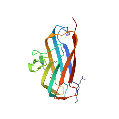
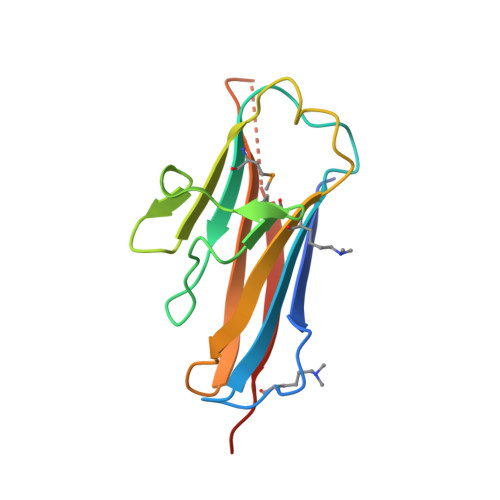
Bsla is a Self-Assembling Bacterial Hydrophobin that Coats the Bacillus Subtilis Biofilm.

Hobley, L.Ostrowski, A.Rao, F.V.Bromley, K.M.Porter, M.Prescott, A.R.Macphee, C.E.Van Aalten, D.M.F.Stanley-Wall, N.R.

(2013) Proc Natl Acad Sci U S A 110: 13600

  • DOI: https://doi.org/10.1073/pnas.1306390110
  • Primary Citation of Related Structures:  
    4BHU

  • PubMed Abstract: 

    Biofilms represent the predominant mode of microbial growth in the natural environment. Bacillus subtilis is a ubiquitous Gram-positive soil bacterium that functions as an effective plant growth-promoting agent. The biofilm matrix is composed of an exopolysaccharide and an amyloid fiber-forming protein, TasA, and assembles with the aid of a small secreted protein, BslA. Here we show that natively synthesized and secreted BslA forms surface layers around the biofilm. Biophysical analysis demonstrates that BslA can self-assemble at interfaces, forming an elastic film. Molecular function is revealed from analysis of the crystal structure of BslA, which consists of an Ig-type fold with the addition of an unusual, extremely hydrophobic "cap" region. A combination of in vivo biofilm formation and in vitro biophysical analysis demonstrates that the central hydrophobic residues of the cap are essential to allow a hydrophobic, nonwetting biofilm to form as they control the surface activity of the BslA protein. The hydrophobic cap exhibits physiochemical properties remarkably similar to the hydrophobic surface found in fungal hydrophobins; thus, BslA is a structurally defined bacterial hydrophobin. We suggest that biofilms formed by other species of bacteria may have evolved similar mechanisms to provide protection to the resident bacterial community.


  • Organizational Affiliation

    Division of Molecular Microbiology, College of Life Sciences, University of Dundee, Dundee DD1 5EH, United Kingdom.


Macromolecules
Find similar proteins by:  (by identity cutoff)  |  3D Structure
Entity ID: 1
MoleculeChains Sequence LengthOrganismDetailsImage
UNCHARACTERIZED PROTEIN YUAB
A, B, C, D, E
A, B, C, D, E, F, G, H, J
130Bacillus subtilis subsp. subtilis NCIB 3610 = ATCC 6051 = DSM 10Mutation(s): 1 
UniProt
Find proteins for P71014 (Bacillus subtilis (strain 168))
Explore P71014 
Go to UniProtKB:  P71014
Entity Groups  
Sequence Clusters30% Identity50% Identity70% Identity90% Identity95% Identity100% Identity
UniProt GroupP71014
Sequence Annotations
Expand
  • Reference Sequence
Find similar proteins by:  (by identity cutoff)  |  3D Structure
Entity ID: 2
MoleculeChains Sequence LengthOrganismDetailsImage
UNCHARACTERIZED PROTEIN YUAB130Bacillus subtilis subsp. subtilis NCIB 3610 = ATCC 6051 = DSM 10Mutation(s): 1 
UniProt
Find proteins for P71014 (Bacillus subtilis (strain 168))
Explore P71014 
Go to UniProtKB:  P71014
Entity Groups  
Sequence Clusters30% Identity50% Identity70% Identity90% Identity95% Identity100% Identity
UniProt GroupP71014
Sequence Annotations
Expand
  • Reference Sequence
Small Molecules
Ligands 2 Unique
IDChains Name / Formula / InChI Key2D Diagram3D Interactions
GOL
Query on GOL

Download Ideal Coordinates CCD File 
K [auth A]
M [auth B]
Q [auth E]
T [auth F]
U [auth F]
K [auth A],
M [auth B],
Q [auth E],
T [auth F],
U [auth F],
Z [auth J]
GLYCEROL
C3 H8 O3
PEDCQBHIVMGVHV-UHFFFAOYSA-N
CL
Query on CL

Download Ideal Coordinates CCD File 
L [auth A]
N [auth B]
O [auth C]
P [auth C]
R [auth E]
L [auth A],
N [auth B],
O [auth C],
P [auth C],
R [auth E],
S [auth E],
V [auth F],
W [auth H],
X [auth I],
Y [auth I]
CHLORIDE ION
Cl
VEXZGXHMUGYJMC-UHFFFAOYSA-M
Modified Residues  2 Unique
IDChains TypeFormula2D DiagramParent
MLY
Query on MLY
A, B, C, D, E
A, B, C, D, E, F, G, H, J
L-PEPTIDE LINKINGC8 H18 N2 O2LYS
MSE
Query on MSE
A, B, C, D, E
A, B, C, D, E, F, G, H, J
L-PEPTIDE LINKINGC5 H11 N O2 SeMET
Experimental Data & Validation

Experimental Data

  • Method: X-RAY DIFFRACTION
  • Resolution: 1.91 Å
  • R-Value Free: 0.199 
  • R-Value Work: 0.165 
  • R-Value Observed: 0.167 
  • Space Group: P 21 21 21
Unit Cell:
Length ( Å )Angle ( ˚ )
a = 69.7α = 90
b = 95.98β = 90
c = 259.81γ = 90
Software Package:
Software NamePurpose
PHENIXrefinement
XDSdata reduction
SCALAdata scaling
PHENIXphasing

Structure Validation

View Full Validation Report


Entry History 

Revision History  (Full details and data files)

  • Version 1.0: 2013-08-14
    Type: Initial release
  • Version 1.1: 2013-08-28
    Changes: Database references