4BBA

Crystal structure of glucokinase regulatory protein complexed to phosphate


Experimental Data Snapshot

  • Method: X-RAY DIFFRACTION
  • Resolution: 1.92 Å
  • R-Value Free: 0.186 
  • R-Value Work: 0.160 
  • R-Value Observed: 0.162 

wwPDB Validation   3D Report Full Report


This is version 1.3 of the entry. See complete history


Literature

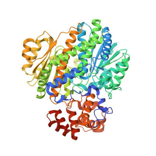
Crystal Structure of Glucokinase Regulatory Protein.

Pautsch, A.Stadler, N.Loehle, A.Rist, W.Berg, A.Glocker, L.Nar, H.Reinert, D.Lenter, M.Heckel, A.Schnapp, G.Kauschke, S.G.

(2013) Biochemistry 52: 3523

  • DOI: https://doi.org/10.1021/bi4000782
  • Primary Citation of Related Structures:  
    4BB9, 4BBA

  • PubMed Abstract: 

    Glucokinase (GK) plays a major role in the regulation of blood glucose homeostasis in both the liver and the pancreas. In the liver, GK is controlled by the GK regulatory protein (GKRP). GKRP in turn is activated by fructose 6-phosphate (F6P) and inactivated by fructose 1-phosphate (F1P). Disrupting the GK-GKRP complex increases the activity of GK in the cytosol and is considered an attractive concept for the regulation of blood glucose. We have determined the crystal structure of GKRP in its inactive F1P-bound form. The binding site for F1P is located deeply buried at a domain interface, and H-D exchange experiments confirmed that F1P and F6P compete for this site. The structure of the inactive GKRP-F1P complex provides a starting point for understanding the mechanism of fructose phosphate-dependent GK regulation at an atomic level.


  • Organizational Affiliation

    Departments of Lead Identification and Optimization Support, ‡CardioMetabolic Diseases Research, §CNS Diseases Research, ∥Drug Discovery Support, ⊥BP Process Science, and @Medicinal Chemistry, Boehringer Ingelheim Pharma GmbH & Company KG , Biberach an der Riss, Germany.


Macromolecules
Find similar proteins by:  (by identity cutoff)  |  3D Structure
Entity ID: 1
MoleculeChains Sequence LengthOrganismDetailsImage
GLUCOKINASE REGULATORY PROTEIN633Homo sapiensMutation(s): 2 
UniProt & NIH Common Fund Data Resources
Find proteins for Q14397 (Homo sapiens)
Explore Q14397 
Go to UniProtKB:  Q14397
PHAROS:  Q14397
Entity Groups  
Sequence Clusters30% Identity50% Identity70% Identity90% Identity95% Identity100% Identity
UniProt GroupQ14397
Sequence Annotations
Expand
  • Reference Sequence
Small Molecules
Ligands 1 Unique
IDChains Name / Formula / InChI Key2D Diagram3D Interactions
PO4
Query on PO4

Download Ideal Coordinates CCD File 
B [auth A]PHOSPHATE ION
O4 P
NBIIXXVUZAFLBC-UHFFFAOYSA-K
Experimental Data & Validation

Experimental Data

  • Method: X-RAY DIFFRACTION
  • Resolution: 1.92 Å
  • R-Value Free: 0.186 
  • R-Value Work: 0.160 
  • R-Value Observed: 0.162 
  • Space Group: P 21 21 21
Unit Cell:
Length ( Å )Angle ( ˚ )
a = 60.76α = 90
b = 72.2β = 90
c = 137.96γ = 90
Software Package:
Software NamePurpose
BUSTERrefinement
XDSdata reduction
XSCALEdata scaling

Structure Validation

View Full Validation Report


Entry History 

Deposition Data

Revision History  (Full details and data files)

  • Version 1.0: 2013-05-15
    Type: Initial release
  • Version 1.1: 2013-08-28
    Changes: Database references
  • Version 1.2: 2019-04-03
    Changes: Data collection, Other, Source and taxonomy
  • Version 1.3: 2023-12-20
    Changes: Data collection, Database references, Derived calculations, Other, Refinement description