4AN4

Crystal structure of the glycosyltransferase SnogD from Streptomyces nogalater


Experimental Data Snapshot

  • Method: X-RAY DIFFRACTION
  • Resolution: 2.70 Å
  • R-Value Free: 0.266 
  • R-Value Work: 0.227 
  • R-Value Observed: 0.229 

wwPDB Validation   3D Report Full Report


Ligand Structure Quality Assessment 


This is version 1.2 of the entry. See complete history


Literature

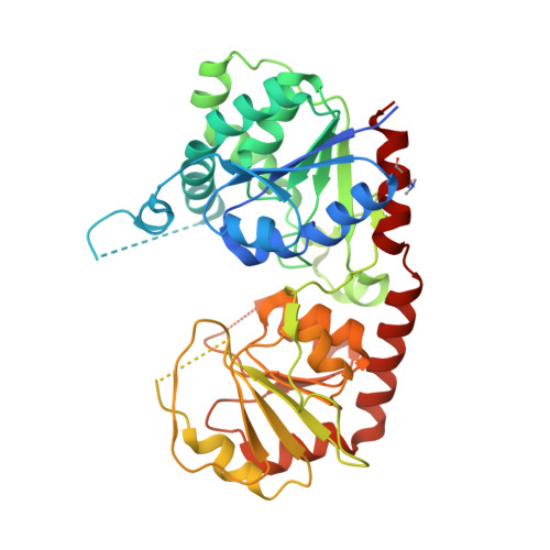
Crystal Structure of the Glycosyltransferase Snogd from the Biosynthetic Pathway of the Nogalamycin in Streptomyces Nogalater.

Claesson, M.Siitonen, V.Dobritzsch, D.Metsa-Ketela, M.Schneider, G.

(2012) FEBS J 279: 3251

  • DOI: https://doi.org/10.1111/j.1742-4658.2012.08711.x
  • Primary Citation of Related Structures:  
    4AMB, 4AMG, 4AN4

  • PubMed Abstract: 

    The glycosyltransferase SnogD from Streptomyces nogalater transfers a nogalamine moiety to the metabolic intermediate 3',4'-demethoxynogalose-1-hydroxynogalamycinone during the final steps of biosynthesis of the aromatic polyketide nogalamycin. The crystal structure of recombinant SnogD, as an apo-enzyme and with a bound nucleotide, 2-deoxyuridine-5'-diphosphate, was determined to 2.6 Å resolution. Reductive methylation of SnogD was crucial for reproducible preparation of diffraction quality crystals due to creation of an additional intermolecular salt bridge between methylated lysine residue Lys384 and Glu374* from an adjacent molecule in the crystal lattice. SnogD is a dimer both in solution and in the crystal, and the enzyme subunit displays a fold characteristic of the GT-B family of glycosyltransferases. Binding of the nucleotide is associated with rearrangement of two active-site loops. Site-directed mutagenesis shows that two active-site histidine residues, His25 and His301, are critical for the glycosyltransferase activities of SnogD both in vivo and in vitro. The crystal structures and the functional data are consistent with a role for His301 in binding of the diphosphate group of the sugar donor substrate, and a function of His25 as a catalytic base in the glycosyl transfer reaction.


  • Organizational Affiliation

    Department of Medical Biochemistry and Biophysics, Karolinska Institutet, Stockholm, Sweden.


Macromolecules
Find similar proteins by:  (by identity cutoff)  |  3D Structure
Entity ID: 1
MoleculeChains Sequence LengthOrganismDetailsImage
GLYCOSYL TRANSFERASE
A, B, C, D
400Streptomyces nogalaterMutation(s): 0 
UniProt
Find proteins for Q9RN61 (Streptomyces nogalater)
Explore Q9RN61 
Go to UniProtKB:  Q9RN61
Entity Groups  
Sequence Clusters30% Identity50% Identity70% Identity90% Identity95% Identity100% Identity
UniProt GroupQ9RN61
Sequence Annotations
Expand
  • Reference Sequence
Small Molecules
Ligands 1 Unique
IDChains Name / Formula / InChI Key2D Diagram3D Interactions
DUD
Query on DUD

Download Ideal Coordinates CCD File 
E [auth B],
F [auth D]
DEOXYURIDINE-5'-DIPHOSPHATE
C9 H14 N2 O11 P2
QHWZTVCCBMIIKE-SHYZEUOFSA-N
Modified Residues  1 Unique
IDChains TypeFormula2D DiagramParent
MLY
Query on MLY
A, B, C, D
L-PEPTIDE LINKINGC8 H18 N2 O2LYS
Experimental Data & Validation

Experimental Data

  • Method: X-RAY DIFFRACTION
  • Resolution: 2.70 Å
  • R-Value Free: 0.266 
  • R-Value Work: 0.227 
  • R-Value Observed: 0.229 
  • Space Group: P 1 2 1
Unit Cell:
Length ( Å )Angle ( ˚ )
a = 64.641α = 90
b = 70.078β = 91.71
c = 176.031γ = 90
Software Package:
Software NamePurpose
REFMACrefinement
MOSFLMdata reduction
SCALAdata scaling
PHASERphasing

Structure Validation

View Full Validation Report


Ligand Structure Quality Assessment 


Entry History 

Deposition Data

Revision History  (Full details and data files)

  • Version 1.0: 2012-09-05
    Type: Initial release
  • Version 1.1: 2018-10-24
    Changes: Data collection, Source and taxonomy
  • Version 1.2: 2023-12-20
    Changes: Data collection, Database references, Derived calculations, Other, Refinement description