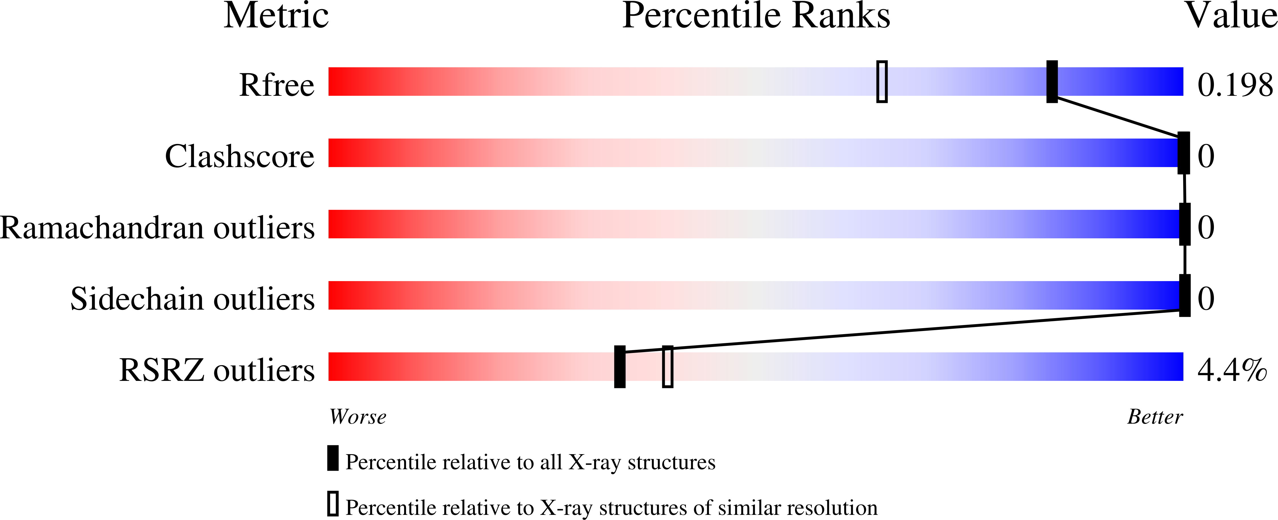
4QT7

Crystal structure of the c-Src SH3 domain in complex with a peptide from the Hepatitis C virus NS5A-protein


Experimental Data Snapshot

  • Method: X-RAY DIFFRACTION
  • Resolution: 1.55 Å
  • R-Value Free: 0.195 
  • R-Value Work: 0.159 
  • R-Value Observed: 0.161 

wwPDB Validation   3D Report Full Report


This is version 1.2 of the entry. See complete history


Literature

Structure of the c-Src-SH3 domain in complex with a proline-rich motif of NS5A protein from the hepatitis C virus.

Bacarizo, J.Martinez-Rodriguez, S.Camara-Artigas, A.

(2015) J Struct Biol 189: 67-72

  • DOI: https://doi.org/10.1016/j.jsb.2014.11.004
  • Primary Citation of Related Structures:  
    4QT7

  • PubMed Abstract: 

    The non-structural hepatitis C virus proteins NS5A and NS5B form a complex through interaction with the SH2 and SH3 domains of the non-receptor Src tyrosine kinase, which seems essential for viral replication. We have crystallized the complex between the SH3 domain of the c-Src tyrosine kinase and the C-terminal proline rich motif of the NS5A protein (A349PPIPPPRRKR359). Crystals obtained at neutral pH belong to the space group I41, with a single molecule of the SH3/NS5A complex at the asymmetric unit. The NS5A peptide is bound in a reverse orientation (class II) and the comparison of this structure with those of the high affinity synthetic peptides APP12 and VSL12 shows some important differences at the salt bridge that drives the peptide orientation. Further conformational changes in residues placed apart from the binding site also seem to play an important role in the binding orientation of this peptide. Our results show the interaction of the SH3 domain of the c-Src tyrosine kinase with a proline rich motif in the NS5A protein and point to their potential interaction in vivo.


  • Organizational Affiliation

    Department of Chemistry and Physics, University of Almería, Agrifood Campus of International Excellence (ceiA3), Carretera de Sacramento s/n, Almería 04120, Spain.


Macromolecules
Find similar proteins by:  (by identity cutoff)  |  3D Structure
Entity ID: 1
MoleculeChains Sequence LengthOrganismDetailsImage
Proto-oncogene tyrosine-protein kinase Src61Gallus gallusMutation(s): 0 
Gene Names: SRC
EC: 2.7.10.2
UniProt
Find proteins for P00523 (Gallus gallus)
Explore P00523 
Go to UniProtKB:  P00523
Entity Groups  
Sequence Clusters30% Identity50% Identity70% Identity90% Identity95% Identity100% Identity
UniProt GroupP00523
Sequence Annotations
Expand
  • Reference Sequence

Find similar proteins by:  Sequence   |   3D Structure  

Entity ID: 2
MoleculeChains Sequence LengthOrganismDetailsImage
NS5A12Hepacivirus hominisMutation(s): 1 
UniProt
Find proteins for V5RF47 (Hepacivirus hominis)
Explore V5RF47 
Go to UniProtKB:  V5RF47
Entity Groups  
Sequence Clusters30% Identity50% Identity70% Identity90% Identity95% Identity100% Identity
UniProt GroupV5RF47
Sequence Annotations
Expand
  • Reference Sequence
Experimental Data & Validation

Experimental Data

  • Method: X-RAY DIFFRACTION
  • Resolution: 1.55 Å
  • R-Value Free: 0.195 
  • R-Value Work: 0.159 
  • R-Value Observed: 0.161 
  • Space Group: I 41
Unit Cell:
Length ( Å )Angle ( ˚ )
a = 62.307α = 90
b = 62.307β = 90
c = 27.266γ = 90
Software Package:
Software NamePurpose
Aimlessdata scaling
PHASERphasing
PHENIXrefinement
PDB_EXTRACTdata extraction
ADSCdata collection
XSCALEdata scaling

Structure Validation

View Full Validation Report


Entry History 

Deposition Data

Revision History  (Full details and data files)

  • Version 1.0: 2014-12-24
    Type: Initial release
  • Version 1.1: 2022-08-24
    Changes: Database references, Derived calculations
  • Version 1.2: 2023-11-08
    Changes: Data collection, Refinement description