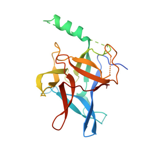
Type 2 Ryanodine Receptor Domain A Contains a Unique and Dynamic alpha-Helix That Transitions to a beta-Strand in a Mutant Linked with a Heritable Cardiomyopathy.
Amador, F.J., Kimlicka, L., Stathopulos, P.B., Gasmi-Seabrook, G.M., Maclennan, D.H., Van Petegem, F., Ikura, M.(2013) J Mol Biol 425: 4034-4046
- PubMed: 23978697
- DOI: https://doi.org/10.1016/j.jmb.2013.08.015
- Primary Citation of Related Structures:
2MC2, 4KEI, 4KEJ, 4KEK - PubMed Abstract:
Ryanodine receptors (RyRs) are large tetrameric calcium (Ca(2+)) release channels found on the sarcoplasmic reticulum that respond to dihydropyridine receptor activity through a direct conformational interaction and/or indirect Ca(2+) sensitivity, propagating sarcoplasmic reticulum luminal Ca(2+) release in the process of excitation-contraction coupling. There are three human RyR subtypes, and several debilitating diseases are linked to heritable mutations in RyR1 and RyR2 including malignant hypothermia, central core disease, catecholaminergic polymorphic ventricular tachycardia (CPVT) and arrhythmogenic right ventricular dysplasia type 2 (ARVD2). Despite the recent appreciation that many disease-associated mutations within the N-terminal RyRABC domains (i.e., residues 1-559) are located in the putative interfaces mediating tetrameric channel assembly, the precise structural and dynamical consequences of the mutations are not well understood. We used solution nuclear magnetic resonance (NMR) spectroscopy and X-ray crystallography to examine the effect of ARVD2-associated (i.e., R176Q) and CPVT-associated [i.e., P164S, R169Q and delta exon 3 (Δ3)] mutations on the structure and dynamics of RyR2A. Our solution NMR data exposed a mobile α-helix, unique to type 2; further, this α2 helix rescues the β-strand lost in RyR2A Δ3 but remains dynamic in the hot-spot loop (HS-loop) P164S, R169Q and R176Q mutant proteins. Docking of our X-ray crystal/NMR hybrid structure into the RyR1 cryo-electron microscopy map revealed that this RyR2A α2 helix is in close proximity to dense "columns" projecting toward the channel pore. This is in contrast to the HS-loop mutations that cause structural changes largely localized to the intersubunit interface between adjacent ABC domains. Taken together, our data suggest that ARVD2 and CPVT mutations have at least two distinct structural consequences linked to channel dysfunction: perturbation of the HS-loop (i.e., domain A):domain B intersubunit interface and disruption of the communication between the N-terminal region and the channel domain.
Organizational Affiliation:
Campbell Family Institute for Cancer Research, Ontario Cancer Institute and Department of Medical Biophysics, University of Toronto, Toronto, Ontario, Canada M5G 1L7.