4JNT

Crystal structure of the ectodomain of Bovine viral diarrhea virus 1 E2 envelope protein


Experimental Data Snapshot

  • Method: X-RAY DIFFRACTION
  • Resolution: 4.09 Å
  • R-Value Free: 0.376 
  • R-Value Work: 0.321 
  • R-Value Observed: 0.323 

wwPDB Validation   3D Report Full Report


This is version 2.1 of the entry. See complete history


Literature

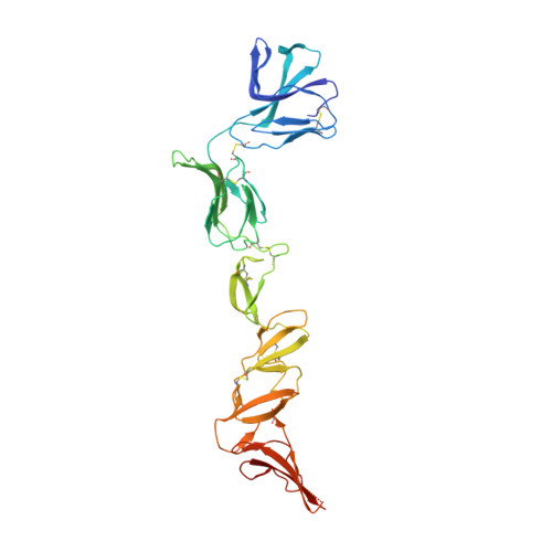
Crystal structure of glycoprotein E2 from bovine viral diarrhea virus.

Li, Y.Wang, J.Kanai, R.Modis, Y.

(2013) Proc Natl Acad Sci U S A 110: 6805-6810

  • DOI: https://doi.org/10.1073/pnas.1300524110
  • Primary Citation of Related Structures:  
    4ILD, 4JNT

  • PubMed Abstract: 

    Pestiviruses, including bovine viral diarrhea virus, are important animal pathogens and are closely related to hepatitis C virus, which remains a major global health threat. They have an outer lipid envelope bearing two glycoproteins, E1 and E2, required for cell entry. They deliver their genome into the host cell cytoplasm by fusion of their envelope with a cellular membrane. The crystal structure of bovine viral diarrhea virus E2 reveals a unique protein architecture consisting of two Ig-like domains followed by an elongated β-stranded domain with a new fold. E2 forms end-to-end homodimers with a conserved C-terminal motif rich in aromatic residues at the contact. A disulfide bond across the interface explains the acid resistance of pestiviruses and their requirement for a redox activation step to initiate fusion. From the structure of E2, we propose alternative possible membrane fusion mechanisms. We expect the pestivirus fusion apparatus to be conserved in hepatitis C virus.


  • Organizational Affiliation

    Department of Molecular Biophysics and Biochemistry, Yale University, New Haven, CT 06520, USA.


Macromolecules
Find similar proteins by:  (by identity cutoff)  |  3D Structure
Entity ID: 1
MoleculeChains Sequence LengthOrganismDetailsImage
Envelope glycoprotein E2
A, B
338Bovine viral diarrhea virus 1Mutation(s): 1 
UniProt
Find proteins for P19711 (Bovine viral diarrhea virus (isolate NADL))
Explore P19711 
Go to UniProtKB:  P19711
Entity Groups  
Sequence Clusters30% Identity50% Identity70% Identity90% Identity95% Identity100% Identity
UniProt GroupP19711
Sequence Annotations
Expand
  • Reference Sequence
Oligosaccharides

Help

Entity ID: 2
MoleculeChains Length2D Diagram Glycosylation3D Interactions
2-acetamido-2-deoxy-beta-D-glucopyranose-(1-4)-2-acetamido-2-deoxy-beta-D-glucopyranose
C, D, E, F, G
C, D, E, F, G, H, I, J
2N-Glycosylation
Glycosylation Resources
GlyTouCan:  G42666HT
GlyCosmos:  G42666HT
GlyGen:  G42666HT
Experimental Data & Validation

Experimental Data

Unit Cell:
Length ( Å )Angle ( ˚ )
a = 155.632α = 90
b = 67.885β = 121.89
c = 139.406γ = 90
Software Package:
Software NamePurpose
HKL-2000data collection
PHASERphasing
REFMACrefinement
XDSdata reduction
XDSdata scaling

Structure Validation

View Full Validation Report


Entry History 

Deposition Data

Revision History  (Full details and data files)

  • Version 1.0: 2013-04-17
    Type: Initial release
  • Version 1.1: 2013-05-22
    Changes: Database references
  • Version 2.0: 2020-07-29
    Type: Remediation
    Reason: Carbohydrate remediation
    Changes: Advisory, Atomic model, Data collection, Database references, Derived calculations, Structure summary
  • Version 2.1: 2023-09-20
    Changes: Data collection, Database references, Refinement description, Structure summary