4G3F

Crystal structure of murine NF-kappaB inducing kinase (NIK) bound to a 2-(aminothiazoly)phenol (cmp2)


Experimental Data Snapshot

  • Method: X-RAY DIFFRACTION
  • Resolution: 1.64 Å
  • R-Value Free: 0.208 
  • R-Value Work: 0.177 
  • R-Value Observed: 0.180 

wwPDB Validation   3D Report Full Report


Ligand Structure Quality Assessment 


This is version 1.3 of the entry. See complete history


Literature

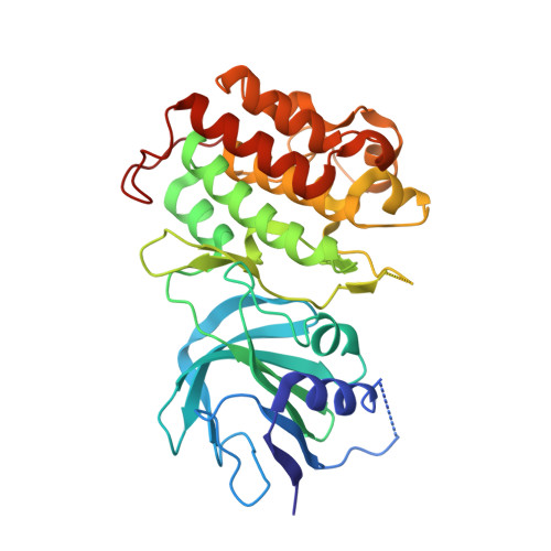
The crystal structure of the catalytic domain of the NF-kappaB inducing kinase reveals a narrow but flexible active site.

de Leon-Boenig, G.Bowman, K.K.Feng, J.A.Crawford, T.Everett, C.Franke, Y.Oh, A.Stanley, M.Staben, S.T.Starovasnik, M.A.Wallweber, H.J.Wu, J.Wu, L.C.Johnson, A.R.Hymowitz, S.G.

(2012) Structure 20: 1704-1714

  • DOI: https://doi.org/10.1016/j.str.2012.07.013
  • Primary Citation of Related Structures:  
    4G3C, 4G3D, 4G3E, 4G3F, 4G3G

  • PubMed Abstract: 

    The NF-κB inducing kinase (NIK) regulates the non-canonical NF-κB pathway downstream of important clinical targets including BAFF, RANKL, and LTβ. Despite numerous genetic studies associating dysregulation of this pathway with autoimmune diseases and hematological cancers, detailed molecular characterization of this central signaling node has been lacking. We undertook a systematic cloning and expression effort to generate soluble, well-behaved proteins encompassing the kinase domains of human and murine NIK. Structures of the apo NIK kinase domain from both species reveal an active-like conformation in the absence of phosphorylation. ATP consumption and peptide phosphorylation assays confirm that phosphorylation of NIK does not increase enzymatic activity. Structures of murine NIK bound to inhibitors possessing two different chemotypes reveal conformational flexibility in the gatekeeper residue controlling access to a hydrophobic pocket. Finally, a single amino acid difference affects the ability of some inhibitors to bind murine and human NIK with the same affinity.


  • Organizational Affiliation

    Department of Structural Biology, Genentech, Inc., 1 DNA Way, South San Francisco, CA 94080, USA.


Macromolecules
Find similar proteins by:  (by identity cutoff)  |  3D Structure
Entity ID: 1
MoleculeChains Sequence LengthOrganismDetailsImage
NF-kappa-beta-inducing kinase336Mus musculusMutation(s): 0 
Gene Names: Map3k14NF-kappaB inducing kinase (NIK)Nik
EC: 2.7.11.25
UniProt
Find proteins for Q9WUL6 (Mus musculus)
Explore Q9WUL6 
Go to UniProtKB:  Q9WUL6
Entity Groups  
Sequence Clusters30% Identity50% Identity70% Identity90% Identity95% Identity100% Identity
UniProt GroupQ9WUL6
Sequence Annotations
Expand
  • Reference Sequence
Small Molecules
Ligands 1 Unique
IDChains Name / Formula / InChI Key2D Diagram3D Interactions
0WB
Query on 0WB

Download Ideal Coordinates CCD File 
B [auth A]3-{2-[(5-fluoro-2-hydroxyphenyl)amino]-1,3-thiazol-4-yl}benzonitrile
C16 H10 F N3 O S
WLSORBAAVBPTGJ-UHFFFAOYSA-N
Binding Affinity Annotations 
IDSourceBinding Affinity
0WB PDBBind:  4G3F Ki: 4.4 (nM) from 1 assay(s)
Experimental Data & Validation

Experimental Data

  • Method: X-RAY DIFFRACTION
  • Resolution: 1.64 Å
  • R-Value Free: 0.208 
  • R-Value Work: 0.177 
  • R-Value Observed: 0.180 
  • Space Group: C 2 2 21
Unit Cell:
Length ( Å )Angle ( ˚ )
a = 74.789α = 90
b = 135.619β = 90
c = 64.857γ = 90
Software Package:
Software NamePurpose
BOSdata collection
PHASERphasing
PHENIXrefinement
HKL-2000data reduction
HKL-2000data scaling

Structure Validation

View Full Validation Report


Ligand Structure Quality Assessment 


Entry History 

Deposition Data

Revision History  (Full details and data files)

  • Version 1.0: 2012-08-08
    Type: Initial release
  • Version 1.1: 2013-01-23
    Changes: Database references
  • Version 1.2: 2018-01-24
    Changes: Structure summary
  • Version 1.3: 2024-02-28
    Changes: Data collection, Database references, Derived calculations