4ARV

Yersinia kristensenii phytase apo form


Experimental Data Snapshot

  • Method: X-RAY DIFFRACTION
  • Resolution: 1.67 Å
  • R-Value Free: 0.198 
  • R-Value Work: 0.164 
  • R-Value Observed: 0.165 

wwPDB Validation   3D Report Full Report


Ligand Structure Quality Assessment 


This is version 1.3 of the entry. See complete history


Literature

Degradation of Phytate by the 6-Phytase from Hafnia Alvei: A Combined Structural and Solution Study.

Ariza, A.Moroz, O.V.Blagova, E.V.Turkenburg, J.P.Waterman, J.Roberts, S.M.Vind, J.Sjoholm, C.Lassen, S.F.De Maria, L.Glitsoe, V.Skov, L.K.Wilson, K.S.

(2013) PLoS One 8: 65062

  • DOI: https://doi.org/10.1371/journal.pone.0065062
  • Primary Citation of Related Structures:  
    4ARO, 4ARS, 4ARU, 4ARV

  • PubMed Abstract: 

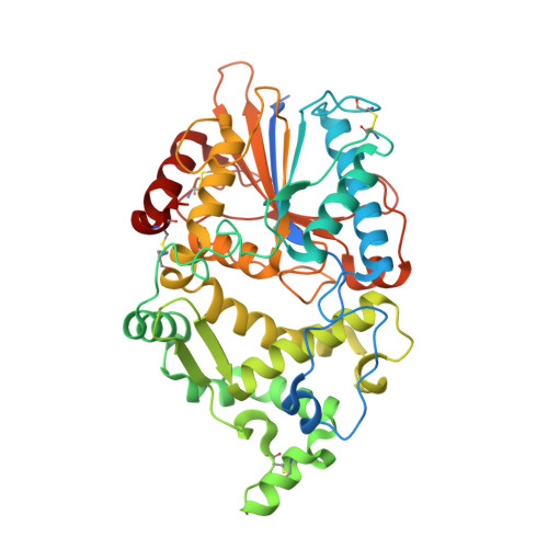
    Phytases hydrolyse phytate (myo-inositol hexakisphosphate), the principal form of phosphate stored in plant seeds to produce phosphate and lower phosphorylated myo-inositols. They are used extensively in the feed industry, and have been characterised biochemically and structurally with a number of structures in the PDB. They are divided into four distinct families: histidine acid phosphatases (HAP), β-propeller phytases, cysteine phosphatases and purple acid phosphatases and also split into three enzyme classes, the 3-, 5- and 6-phytases, depending on the position of the first phosphate in the inositol ring to be removed. We report identification, cloning, purification and 3D structures of 6-phytases from two bacteria, Hafnia alvei and Yersinia kristensenii, together with their pH optima, thermal stability, and degradation profiles for phytate. An important result is the structure of the H. alvei enzyme in complex with the substrate analogue myo-inositol hexakissulphate. In contrast to the only previous structure of a ligand-bound 6-phytase, where the 3-phosphate was unexpectedly in the catalytic site, in the H. alvei complex the expected scissile 6-phosphate (sulphate in the inhibitor) is placed in the catalytic site.


  • Organizational Affiliation

    Structural Biology Laboratory, Department of Chemistry, University of York, York, United Kingdom.


Macromolecules
Find similar proteins by:  (by identity cutoff)  |  3D Structure
Entity ID: 1
MoleculeChains Sequence LengthOrganismDetailsImage
PHYTASE
A, B
441Yersinia kristenseniiMutation(s): 0 
EC: 3.1.3.26
UniProt
Find proteins for H9TUK6 (Yersinia kristensenii)
Explore H9TUK6 
Go to UniProtKB:  H9TUK6
Entity Groups  
Sequence Clusters30% Identity50% Identity70% Identity90% Identity95% Identity100% Identity
UniProt GroupH9TUK6
Sequence Annotations
Expand
  • Reference Sequence
Small Molecules
Ligands 4 Unique
IDChains Name / Formula / InChI Key2D Diagram3D Interactions
P15
Query on P15

Download Ideal Coordinates CCD File 
N [auth B]2,5,8,11,14,17-HEXAOXANONADECAN-19-OL
C13 H28 O7
FHHGCKHKTAJLOM-UHFFFAOYSA-N
TOE
Query on TOE

Download Ideal Coordinates CCD File 
H [auth A]2-[2-(2-METHOXY-ETHOXY)-ETHOXY]-ETHOXYL
C7 H16 O4
JLGLQAWTXXGVEM-UHFFFAOYSA-N
PO4
Query on PO4

Download Ideal Coordinates CCD File 
C [auth A],
D [auth A],
I [auth B],
J [auth B]
PHOSPHATE ION
O4 P
NBIIXXVUZAFLBC-UHFFFAOYSA-K
EDO
Query on EDO

Download Ideal Coordinates CCD File 
E [auth A]
F [auth A]
G [auth A]
K [auth B]
L [auth B]
E [auth A],
F [auth A],
G [auth A],
K [auth B],
L [auth B],
M [auth B]
1,2-ETHANEDIOL
C2 H6 O2
LYCAIKOWRPUZTN-UHFFFAOYSA-N
Experimental Data & Validation

Experimental Data

  • Method: X-RAY DIFFRACTION
  • Resolution: 1.67 Å
  • R-Value Free: 0.198 
  • R-Value Work: 0.164 
  • R-Value Observed: 0.165 
  • Space Group: P 1
Unit Cell:
Length ( Å )Angle ( ˚ )
a = 55.41α = 76.67
b = 67.723β = 87.35
c = 73.164γ = 78.32
Software Package:
Software NamePurpose
REFMACrefinement
MOLREPphasing

Structure Validation

View Full Validation Report


Ligand Structure Quality Assessment 


Entry History 

Revision History  (Full details and data files)

  • Version 1.0: 2013-05-08
    Type: Initial release
  • Version 1.1: 2013-06-19
    Changes: Database references, Non-polymer description
  • Version 1.2: 2014-02-05
    Changes: Database references
  • Version 1.3: 2023-12-20
    Changes: Data collection, Database references, Derived calculations, Other, Refinement description