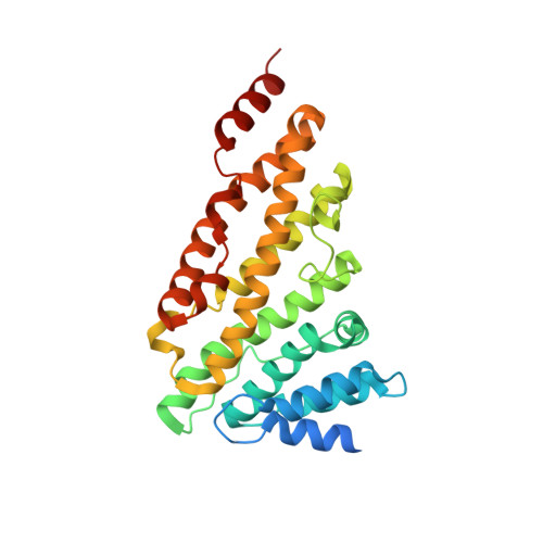
3ZYK

Structure of CALM (PICALM) ANTH domain


Experimental Data Snapshot

  • Method: X-RAY DIFFRACTION
  • Resolution: 1.80 Å
  • R-Value Free: 0.216 
  • R-Value Work: 0.182 
  • R-Value Observed: 0.183 

wwPDB Validation   3D Report Full Report


This is version 1.2 of the entry. See complete history


Literature

The molecular basis for the endocytosis of small R-SNAREs by the clathrin adaptor CALM.

Miller, S.E.Sahlender, D.A.Graham, S.C.Honing, S.Robinson, M.S.Peden, A.A.Owen, D.J.

(2011) Cell 147: 1118-1131

  • DOI: https://doi.org/10.1016/j.cell.2011.10.038
  • Primary Citation of Related Structures:  
    3ZYK, 3ZYL, 3ZYM

  • PubMed Abstract: 

    SNAREs provide a large part of the specificity and energy needed for membrane fusion and, to do so, must be localized to their correct membranes. Here, we show that the R-SNAREs VAMP8, VAMP3, and VAMP2, which cycle between the plasma membrane and endosomes, bind directly to the ubiquitously expressed, PtdIns4,5P(2)-binding, endocytic clathrin adaptor CALM/PICALM. X-ray crystallography shows that the N-terminal halves of their SNARE motifs bind the CALM(ANTH) domain as helices in a manner that mimics SNARE complex formation. Mutation of residues in the CALM:SNARE interface inhibits binding in vitro and prevents R-SNARE endocytosis in vivo. Thus, CALM:R-SNARE interactions ensure that R-SNAREs, required for the fusion of endocytic clathrin-coated vesicles with endosomes and also for subsequent postendosomal trafficking, are sorted into endocytic vesicles. CALM's role in directing the endocytosis of small R-SNAREs may provide insight into the association of CALM/PICALM mutations with growth retardation, cognitive defects, and Alzheimer's disease.


  • Organizational Affiliation

    Cambridge Institute for Medical Research and Department of Clinical Biochemistry, University of Cambridge, Addenbrooke's Hospital, Hills Road, Cambridge CB2 0XY, UK.


Macromolecules
Find similar proteins by:  (by identity cutoff)  |  3D Structure
Entity ID: 1
MoleculeChains Sequence LengthOrganismDetailsImage
PHOSPHATIDYLINOSITOL-BINDING CLATHRIN ASSEMBLY PROTEIN
A, B
296Rattus norvegicusMutation(s): 0 
UniProt
Find proteins for O55012 (Rattus norvegicus)
Explore O55012 
Go to UniProtKB:  O55012
Entity Groups  
Sequence Clusters30% Identity50% Identity70% Identity90% Identity95% Identity100% Identity
UniProt GroupO55012
Sequence Annotations
Expand
  • Reference Sequence
Experimental Data & Validation

Experimental Data

  • Method: X-RAY DIFFRACTION
  • Resolution: 1.80 Å
  • R-Value Free: 0.216 
  • R-Value Work: 0.182 
  • R-Value Observed: 0.183 
  • Space Group: P 21 21 21
Unit Cell:
Length ( Å )Angle ( ˚ )
a = 73.541α = 90
b = 78.313β = 90
c = 120.217γ = 90
Software Package:
Software NamePurpose
PHENIXrefinement
MOSFLMdata reduction
SCALAdata scaling
PHASERphasing

Structure Validation

View Full Validation Report


Entry History 

Deposition Data

Revision History  (Full details and data files)

  • Version 1.0: 2011-12-07
    Type: Initial release
  • Version 1.1: 2019-03-13
    Changes: Data collection, Database references, Experimental preparation, Other
  • Version 1.2: 2023-12-20
    Changes: Data collection, Database references, Other, Refinement description