


Peptidic Alpha, Beta-Unsaturated Ethyl Esters as Inhibitors of the 3C Protease of Coxsackie Virus B3: Crystal Structures, Antiviral Activities, and Resistance Mutations
Tan, J., Anand, K., Mesters, J.R., Hilgenfeld, R.To be published.
Experimental Data Snapshot
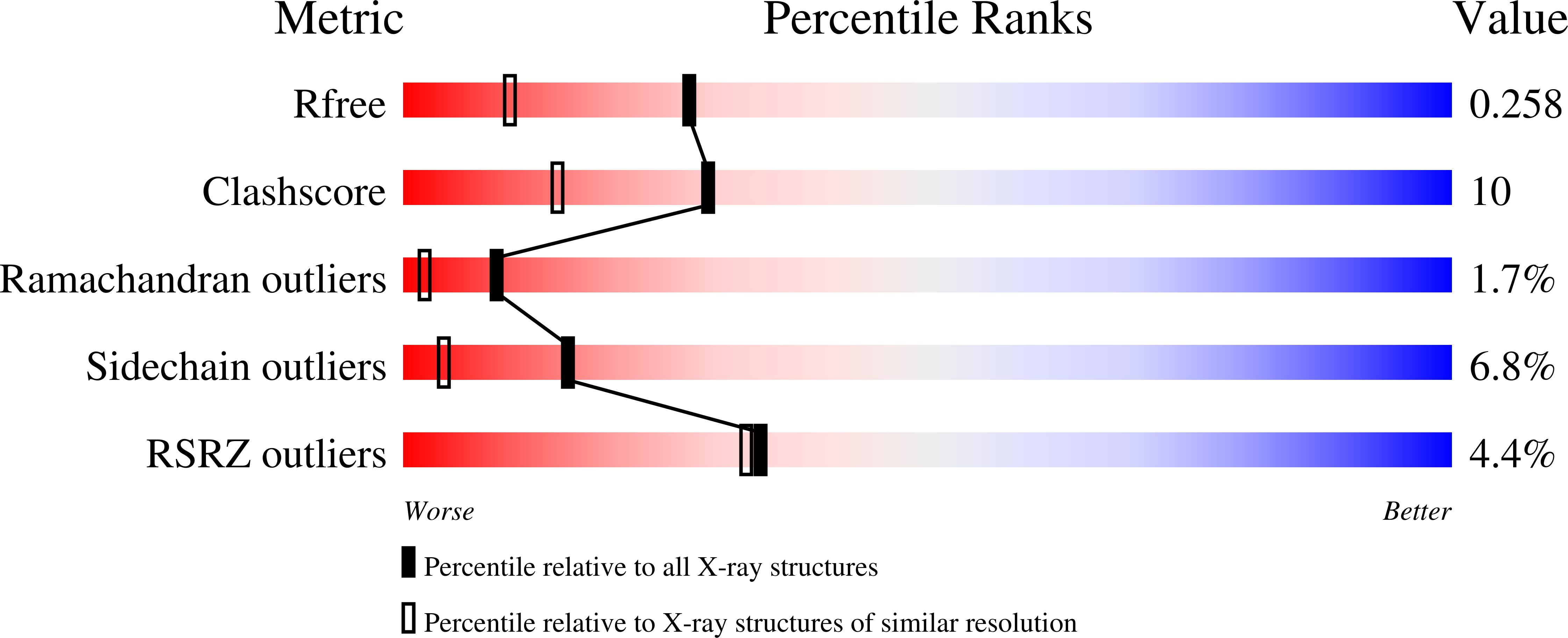
wwPDB Validation 3D Report Full Report
Entity ID: 1 | |||||
|---|---|---|---|---|---|
| Molecule | Chains | Sequence Length | Organism | Details | Image |
| 3C PROTEINASE | 184 | Coxsackievirus B3 | Mutation(s): 1 EC: 3.4.22.28 | 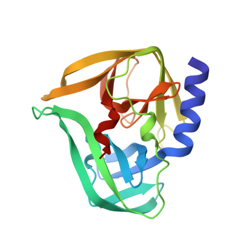 | |
UniProt | |||||
Find proteins for Q5UEA2 (Coxsackievirus B3) Explore Q5UEA2 Go to UniProtKB: Q5UEA2 | |||||
Entity Groups | |||||
| Sequence Clusters | 30% Identity50% Identity70% Identity90% Identity95% Identity100% Identity | ||||
| UniProt Group | Q5UEA2 | ||||
Sequence AnnotationsExpand | |||||
| |||||
| Length ( Å ) | Angle ( ˚ ) |
|---|---|
| a = 76.73 | α = 90 |
| b = 64.44 | β = 115.78 |
| c = 39.81 | γ = 90 |
| Software Name | Purpose |
|---|---|
| REFMAC | refinement |
| iMOSFLM | data reduction |
| SCALA | data scaling |
| MOLREP | phasing |
RCSB PDB (citation) is hosted by
RCSB PDB is a member of the

