3ZM6

CRYSTAL STRUCTURE OF MURF LIGASE IN COMPLEX WITH CYANOTHIOPHENE INHIBITOR


Experimental Data Snapshot

  • Method: X-RAY DIFFRACTION
  • Resolution: 1.84 Å
  • R-Value Free: 0.255 
  • R-Value Work: 0.224 
  • R-Value Observed: 0.227 

wwPDB Validation   3D Report Full Report


Ligand Structure Quality Assessment 


This is version 1.1 of the entry. See complete history


Literature

Structure-Activity Relationships of New Cyanothiophene Inhibitors of the Essential Peptidoglycan Biosynthesis Enzyme Murf.

Hrast, M.Turk, S.Sosic, I.Knez, D.Randall, C.P.Barreteau, H.Contreras-Martel, C.Dessen, A.O'Neill, A.J.Mengin-Lecreulx, D.Blanot, D.Gobec, S.

(2013) Eur J Med Chem 66C: 32

  • DOI: https://doi.org/10.1016/j.ejmech.2013.05.013
  • Primary Citation of Related Structures:  
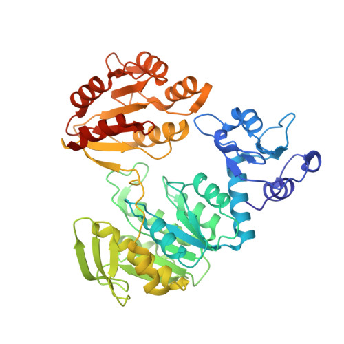
    3ZM5, 3ZM6

  • PubMed Abstract: 

    Peptidoglycan is an essential component of the bacterial cell wall, and enzymes involved in its biosynthesis represent validated targets for antibacterial drug discovery. MurF catalyzes the final intracellular peptidoglycan biosynthesis step: the addition of D-Ala-D-Ala to the nucleotide precursor UDP-MurNAc-L-Ala-γ-D-Glu-meso-DAP (or L-Lys). As MurF has no human counterpart, it represents an attractive target for the development of new antibacterial drugs. Using recently published cyanothiophene inhibitors of MurF from Streptococcus pneumoniae as a starting point, we designed and synthesized a series of structurally related derivatives and investigated their inhibition of MurF enzymes from different bacterial species. Systematic structural modifications of the parent compounds resulted in a series of nanomolar inhibitors of MurF from S. pneumoniae and micromolar inhibitors of MurF from Escherichia coli and Staphylococcus aureus. Some of the inhibitors also show antibacterial activity against S. pneumoniae R6. These findings, together with two new co-crystal structures, represent an excellent starting point for further optimization toward effective novel antibacterials.


  • Organizational Affiliation

    Faculty of Pharmacy, University of Ljubljana, Aškerčeva 7, 1000 Ljubljana, Slovenia.


Macromolecules
Find similar proteins by:  (by identity cutoff)  |  3D Structure
Entity ID: 1
MoleculeChains Sequence LengthOrganismDetailsImage
UDP-N-ACETYLMURAMOYL-TRIPEPTIDE--D-ALANYL-D-ALANINE LIGASE465Streptococcus pneumoniae R6Mutation(s): 0 
EC: 6.3.2.10
UniProt
Find proteins for Q8DNV6 (Streptococcus pneumoniae (strain ATCC BAA-255 / R6))
Explore Q8DNV6 
Go to UniProtKB:  Q8DNV6
Entity Groups  
Sequence Clusters30% Identity50% Identity70% Identity90% Identity95% Identity100% Identity
UniProt GroupQ8DNV6
Sequence Annotations
Expand
  • Reference Sequence
Small Molecules
Ligands 1 Unique
IDChains Name / Formula / InChI Key2D Diagram3D Interactions
2GN
Query on 2GN

Download Ideal Coordinates CCD File 
B [auth A]N-(6-(4-(2h-tetrazol-5-yl)benzyl)-3-cyano-4,5,6,7-tetrahydrothieno[2,3-c]pyridin-2-yl)-2,4-dichloro-5-(morpholinosulfonyl)benzamide
C27 H24 Cl2 N8 O4 S2
KXJHJZRFLLSTJD-UHFFFAOYSA-N
Binding Affinity Annotations 
IDSourceBinding Affinity
2GN Binding MOAD:  3ZM6 IC50: 300 (nM) from 1 assay(s)
PDBBind:  3ZM6 IC50: 300 (nM) from 1 assay(s)
Experimental Data & Validation

Experimental Data

  • Method: X-RAY DIFFRACTION
  • Resolution: 1.84 Å
  • R-Value Free: 0.255 
  • R-Value Work: 0.224 
  • R-Value Observed: 0.227 
  • Space Group: P 61 2 2
Unit Cell:
Length ( Å )Angle ( ˚ )
a = 116.156α = 90
b = 116.156β = 90
c = 160.294γ = 120
Software Package:
Software NamePurpose
REFMACrefinement
XDSdata reduction
XSCALEdata scaling
PHASERphasing

Structure Validation

View Full Validation Report


Ligand Structure Quality Assessment 


Entry History 

Revision History  (Full details and data files)

  • Version 1.0: 2013-07-03
    Type: Initial release
  • Version 1.1: 2023-12-20
    Changes: Data collection, Database references, Derived calculations, Other, Refinement description