3ZGQ

Crystal structure of human interferon-induced protein IFIT5


Experimental Data Snapshot

  • Method: X-RAY DIFFRACTION
  • Resolution: 2.20 Å
  • R-Value Free: 0.240 
  • R-Value Work: 0.178 
  • R-Value Observed: 0.182 

wwPDB Validation   3D Report Full Report


This is version 1.1 of the entry. See complete history


Literature

TRNA Binding, Structure, and Localization of the Human Interferon-Induced Protein Ifit5.

Katibah, G.E.Lee, H.J.Huizar, J.P.Vogan, J.M.Alber, T.Collins, K.

(2013) Mol Cell 49: 743

  • DOI: https://doi.org/10.1016/j.molcel.2012.12.015
  • Primary Citation of Related Structures:  
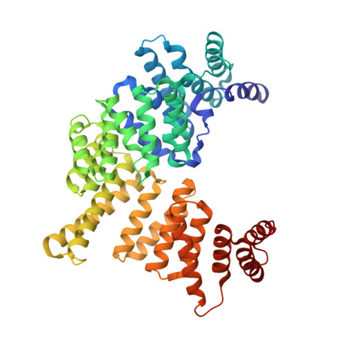
    3ZGQ

  • PubMed Abstract: 

    Interferon-induced proteins, including the largely uncharacterized interferon-induced tetratricopeptide repeat (IFIT) protein family, provide defenses against pathogens. Differing from expectations for tetratricopeptide repeat (TPR) proteins and from human IFIT1, IFIT2, and IFIT3, we show that human IFIT5 recognizes cellular RNA instead of protein partners. In vivo and in vitro, IFIT5 bound to endogenous 5'-phosphate-capped RNAs, including transfer RNAs. The crystal structure of IFIT5 revealed a convoluted intramolecular packing of eight TPRs as a fold that we name the TPR eddy. Additional, non-TPR structural elements contribute to an RNA binding cleft. Instead of general cytoplasmic distribution, IFIT5 concentrated in actin-rich protrusions from the apical cell surface colocalized with the RNA-binding retinoic acid-inducible gene-I (RIG-I). These findings establish compartmentalized cellular RNA binding activity as a mechanism for IFIT5 function and reveal the TPR eddy as a scaffold for RNA recognition.


  • Organizational Affiliation

    Department of Molecular and Cell Biology, University of California, Berkeley, Berkeley, CA 94720, USA.


Macromolecules
Find similar proteins by:  (by identity cutoff)  |  3D Structure
Entity ID: 1
MoleculeChains Sequence LengthOrganismDetailsImage
INTERFERON-INDUCED PROTEIN WITH TETRATRICOPEPTIDE REPEATS 5482Homo sapiensMutation(s): 0 
UniProt & NIH Common Fund Data Resources
Find proteins for Q13325 (Homo sapiens)
Explore Q13325 
Go to UniProtKB:  Q13325
PHAROS:  Q13325
GTEx:  ENSG00000152778 
Entity Groups  
Sequence Clusters30% Identity50% Identity70% Identity90% Identity95% Identity100% Identity
UniProt GroupQ13325
Sequence Annotations
Expand
  • Reference Sequence
Small Molecules
Ligands 1 Unique
IDChains Name / Formula / InChI Key2D Diagram3D Interactions
PEG
Query on PEG

Download Ideal Coordinates CCD File 
B [auth A]DI(HYDROXYETHYL)ETHER
C4 H10 O3
MTHSVFCYNBDYFN-UHFFFAOYSA-N
Experimental Data & Validation

Experimental Data

  • Method: X-RAY DIFFRACTION
  • Resolution: 2.20 Å
  • R-Value Free: 0.240 
  • R-Value Work: 0.178 
  • R-Value Observed: 0.182 
  • Space Group: P 21 21 21
Unit Cell:
Length ( Å )Angle ( ˚ )
a = 63.833α = 90
b = 71.793β = 90
c = 116.92γ = 90
Software Package:
Software NamePurpose
PHENIXrefinement
HKL-2000data reduction
HKL-2000data scaling

Structure Validation

View Full Validation Report


Entry History 

Deposition Data

Revision History  (Full details and data files)

  • Version 1.0: 2013-01-23
    Type: Initial release
  • Version 1.1: 2013-03-06
    Changes: Database references