3ZG1

NI-BOUND FORM OF M123A MUTANT OF CUPRIAVIDUS METALLIDURANS CH34 CNRXS


Experimental Data Snapshot

  • Method: X-RAY DIFFRACTION
  • Resolution: 1.85 Å
  • R-Value Free: 0.222 
  • R-Value Work: 0.176 
  • R-Value Observed: 0.177 

wwPDB Validation   3D Report Full Report


This is version 1.3 of the entry. See complete history


Literature

Metal Sensing and Signal Transduction by Cnrx from Cupriavidus Metallidurans Ch34: Role of the Only Methionine Assessed by a Functional, Spectroscopic, and Theoretical Study

Trepreau, J.Grosse, C.Mouesca, J.-M.Sarret, G.Girard, E.Petit-Haertlein, I.Kuennemann, S.Desbourdes, C.De Rosny, E.Maillard, A.P.Nies, D.H.Coves, J.

(2014) Metallomics 6: 263

  • DOI: https://doi.org/10.1039/c3mt00248a
  • Primary Citation of Related Structures:  
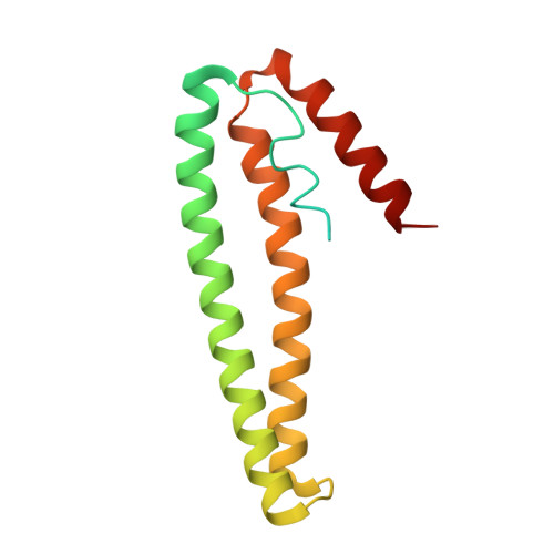
    3ZG1

  • PubMed Abstract: 

    When CnrX, the periplasmic sensor protein in the CnrYXH transmembrane signal transduction complex of Cupriavidus metallidurans CH34, binds the cognate metal ions Ni(II) or Co(II), the ECF-type sigma factor CnrH is made available in the cytoplasm for the RNA-polymerase to initiate transcription at the cnrYp and cnrCp promoters. Ni(II) or Co(II) are sensed by a metal-binding site with a N3O2S coordination sphere with octahedral geometry, where S stands for the thioether sulfur of the only methionine (Met123) residue of CnrX. The M123A-CnrX derivative has dramatically reduced signal propagation in response to metal sensing while the X-ray structure of Ni-bound M123A-CnrXs showed that the metal-binding site was not affected by the mutation. Ni(II) remained six-coordinate in M123A-CnrXs, with a water molecule replacing the sulfur as the sixth ligand. H32A-CnrXs, the soluble model of the wild-type membrane-anchored CnrX, was compared to the double mutants H32A-M123A-CnrXs and H32A-M123C-CnrXs to spectroscopically evaluate the role of this unique ligand in the binding site of Ni or Co. The Co- and Ni-bound forms of the protein display unusually blue-shifted visible spectra. TD-DFT calculations using structure-based models allowed identification and assignment of the electronic transitions of Co-bound form of the protein and its M123A derivative. Among them, the signature of the S-Co transition is distinguishable in the shoulder at 530 nm. In vitro affinity measurements point out the crucial role of Met123 in the selectivity for Ni or Co, and in vivo data support the conclusion that Met123 is a trigger of the signal transduction.


  • Organizational Affiliation

    Institut de Biologie Structurale, UMR 5075 CNRS-CEA-UJF-Grenoble-1, 6 Rue Jules Horowitz, 38042 Grenoble, France. jacques.coves@ibs.fr.


Macromolecules
Find similar proteins by:  (by identity cutoff)  |  3D Structure
Entity ID: 1
MoleculeChains Sequence LengthOrganismDetailsImage
NICKEL AND COBALT RESISTANCE PROTEIN CNRR
A, B, C, D
148Cupriavidus metallidurans CH34Mutation(s): 1 
UniProt
Find proteins for P37975 (Cupriavidus metallidurans (strain ATCC 43123 / DSM 2839 / NBRC 102507 / CH34))
Explore P37975 
Go to UniProtKB:  P37975
Entity Groups  
Sequence Clusters30% Identity50% Identity70% Identity90% Identity95% Identity100% Identity
UniProt GroupP37975
Sequence Annotations
Expand
  • Reference Sequence
Small Molecules
Ligands 5 Unique
IDChains Name / Formula / InChI Key2D Diagram3D Interactions
PGE
Query on PGE

Download Ideal Coordinates CCD File 
Q [auth C]TRIETHYLENE GLYCOL
C6 H14 O4
ZIBGPFATKBEMQZ-UHFFFAOYSA-N
PEG
Query on PEG

Download Ideal Coordinates CCD File 
E [auth A],
G [auth A],
H [auth A],
P [auth C]
DI(HYDROXYETHYL)ETHER
C4 H10 O3
MTHSVFCYNBDYFN-UHFFFAOYSA-N
GOL
Query on GOL

Download Ideal Coordinates CCD File 
L [auth C],
N [auth C],
O [auth C]
GLYCEROL
C3 H8 O3
PEDCQBHIVMGVHV-UHFFFAOYSA-N
NI
Query on NI

Download Ideal Coordinates CCD File 
F [auth A],
K [auth B],
M [auth C],
R [auth D]
NICKEL (II) ION
Ni
VEQPNABPJHWNSG-UHFFFAOYSA-N
CL
Query on CL

Download Ideal Coordinates CCD File 
I [auth B],
J [auth B]
CHLORIDE ION
Cl
VEXZGXHMUGYJMC-UHFFFAOYSA-M
Experimental Data & Validation

Experimental Data

  • Method: X-RAY DIFFRACTION
  • Resolution: 1.85 Å
  • R-Value Free: 0.222 
  • R-Value Work: 0.176 
  • R-Value Observed: 0.177 
  • Space Group: P 1 21 1
Unit Cell:
Length ( Å )Angle ( ˚ )
a = 31.846α = 90
b = 79.125β = 90.1
c = 93.475γ = 90
Software Package:
Software NamePurpose
PHENIXrefinement
XDSdata reduction
Aimlessdata scaling
PHASERphasing

Structure Validation

View Full Validation Report


Entry History 

Deposition Data

Revision History  (Full details and data files)

  • Version 1.0: 2013-10-23
    Type: Initial release
  • Version 1.1: 2013-11-06
    Changes: Database references
  • Version 1.2: 2014-02-12
    Changes: Database references
  • Version 1.3: 2023-12-20
    Changes: Data collection, Database references, Derived calculations, Other, Refinement description