Structural and Catalytic Effects of an Invariant Purine Substitution in the Hammerhead Ribozyme: Implications for the Mechanism of Acid-Base Catalysis.
Schultz, E.P., Vasquez, E.E., Scott, W.G.(2014) Acta Crystallogr D Biol Crystallogr 70: 2256
- PubMed: 25195740
- DOI: https://doi.org/10.1107/S1399004714010608
- Primary Citation of Related Structures:
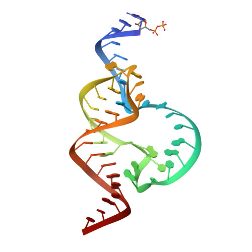
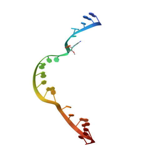
3ZD4 - PubMed Abstract:
The hammerhead ribozyme catalyzes RNA cleavage via acid-base catalysis. Whether it does so by general acid-base catalysis, in which the RNA itself donates and abstracts protons in the transition state, as is typically assumed, or by specific acid-base catalysis, in which the RNA plays a structural role and proton transfer is mediated by active-site water molecules, is unknown. Previous biochemical and crystallographic experiments implicate an invariant purine in the active site, G12, as the general base. However, G12 may play a structural role consistent with specific base catalysis. To better understand the role of G12 in the mechanism of hammerhead catalysis, a 2.2 Å resolution crystal structure of a hammerhead ribozyme from Schistosoma mansoni with a purine substituted for G12 in the active site of the ribozyme was obtained. Comparison of this structure (PDB entry 3zd4), in which A12 is substituted for G, with three previously determined structures that now serve as important experimental controls, allows the identification of structural perturbations that are owing to the purine substitution itself. Kinetic measurements for G12 purine-substituted schistosomal hammerheads confirm a previously observed dependence of rate on the pK(a) of the substituted purine; in both cases inosine, which is similar to G in pK(a) and hydrogen-bonding properties, is unexpectedly inactive. Structural comparisons indicate that this may primarily be owing to the lack of the exocyclic 2-amino group in the G12A and G12I substitutions and its structural effect upon both the nucleotide base and phosphate of A9. The latter involves the perturbation of a previously identified and well characterized metal ion-binding site known to be catalytically important in both minimal and full-length hammerhead ribozyme sequences. The results permit it to be suggested that G12 plays an important role in stabilizing the active-site structure. This result, although not inconsistent with the potential role of G12 as a general base, indicates that an alternative hammerhead cleavage mechanism involving specific base catalysis may instead explain the observed rate dependence upon purine substitutions at G12. The crystallographic results, contrary to previous assumptions, therefore cannot be interpreted to favor the general base catalysis mecahnism over the specific base catalysis mechanism. Instead, both of these mutually exclusive mechanistic alternatives must be considered in light of the current structural and biochemical data.
Organizational Affiliation:
Department of Chemistry and Biochemistry and The Center for the Molecular Biology of RNA, University of California at Santa Cruz, Santa Cruz, CA 95064, USA.