


Crystal structure of wild-type of E. coli CutA1
Tanaka, T., Matsuura, Y., Yutani, K.To be published.
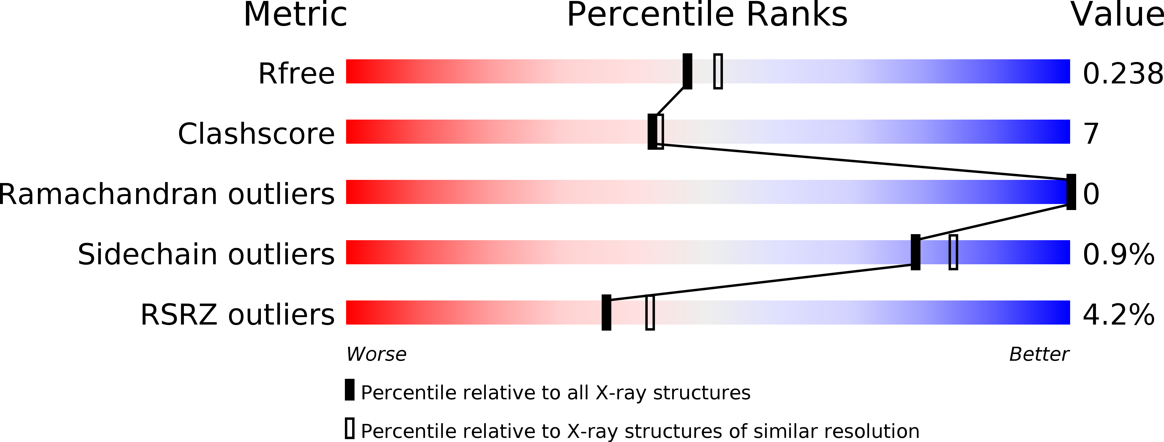
Experimental Data Snapshot
wwPDB Validation 3D Report Full Report
Entity ID: 1 | |||||
|---|---|---|---|---|---|
| Molecule | Chains | Sequence Length | Organism | Details | Image |
| Divalent-cation tolerance protein CutA | 112 | Escherichia coli K-12 | Mutation(s): 0 Gene Names: b4137, cutA, cutA1, cycY, JW4097 | 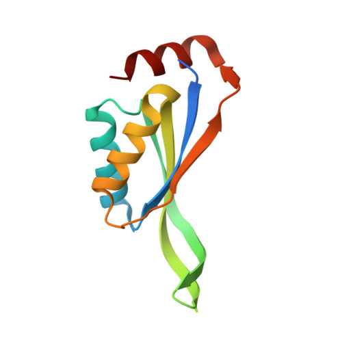 | |
UniProt | |||||
Find proteins for P69488 (Escherichia coli (strain K12)) Explore P69488 Go to UniProtKB: P69488 | |||||
Entity Groups | |||||
| Sequence Clusters | 30% Identity50% Identity70% Identity90% Identity95% Identity100% Identity | ||||
| UniProt Group | P69488 | ||||
Sequence AnnotationsExpand | |||||
| |||||
| Length ( Å ) | Angle ( ˚ ) |
|---|---|
| a = 62.419 | α = 90 |
| b = 96.868 | β = 90 |
| c = 106.398 | γ = 90 |
| Software Name | Purpose |
|---|---|
| HKL-2000 | data collection |
| MOLREP | phasing |
| REFMAC | refinement |
| HKL-2000 | data reduction |
| HKL-2000 | data scaling |
RCSB PDB (citation) is hosted by
RCSB PDB is a member of the

