3WSR

Crystal structure of CLEC-2 in complex with O-glycosylated podoplanin


Experimental Data Snapshot

  • Method: X-RAY DIFFRACTION
  • Resolution: 1.91 Å
  • R-Value Free: 0.259 
  • R-Value Work: 0.217 
  • R-Value Observed: 0.219 

wwPDB Validation   3D Report Full Report


This is version 2.1 of the entry. See complete history


Literature

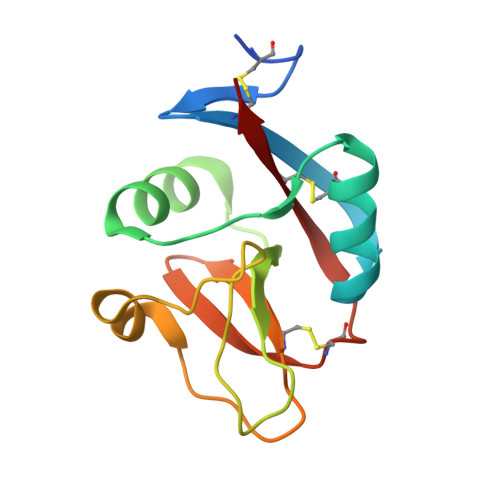
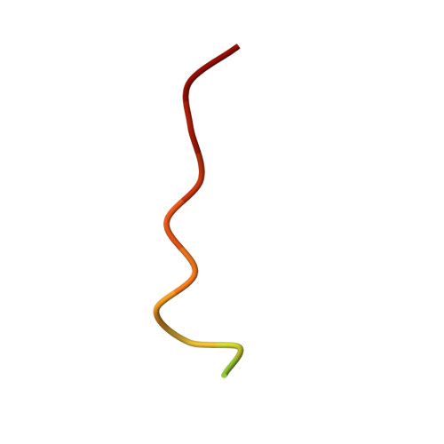
A Platform of C-type Lectin-like Receptor CLEC-2 for Binding O-Glycosylated Podoplanin and Nonglycosylated Rhodocytin

Nagae, M.Morita-Matsumoto, K.Kato, M.Kato-Kaneko, M.Kato, Y.Yamaguchi, Y.

(2014) Structure 22: 1711-1721

  • DOI: https://doi.org/10.1016/j.str.2014.09.009
  • Primary Citation of Related Structures:  
    3WSR, 3WWK

  • PubMed Abstract: 

    Podoplanin is a transmembrane O-glycoprotein that binds to C-type lectin-like receptor 2 (CLEC-2). The O-glycan-dependent interaction seems to play crucial roles in various biological processes, such as platelet aggregation. Rhodocytin, a snake venom, also binds to CLEC-2 and aggregates platelets in a glycan-independent manner. To elucidate the structural basis of the glycan-dependent and independent interactions, we performed comparative crystallographic studies of podoplanin and rhodocytin in complex with CLEC-2. Both podoplanin and rhodocytin bind to the noncanonical "side" face of CLEC-2. There is a common interaction mode between consecutive acidic residues on the ligands and the same arginine residues on CLEC-2. Other interactions are ligand-specific. Carboxyl groups from the sialic acid residue on podoplanin and from the C terminus of the rhodocytin α subunit interact differently at this "second" binding site on CLEC-2. The unique and versatile binding modes open a way to understand the functional consequences of CLEC-2-ligand interactions.


  • Organizational Affiliation

    Structural Glycobiology Team, Systems Glycobiology Research Group, RIKEN-Max Planck Joint Research Center, RIKEN Global Research Cluster, 2-1 Hirosawa, Wako, Saitama 351-0198, Japan.


Macromolecules
Find similar proteins by:  (by identity cutoff)  |  3D Structure
Entity ID: 1
MoleculeChains Sequence LengthOrganismDetailsImage
C-type lectin domain family 1 member B
A, B
128Homo sapiensMutation(s): 1 
Gene Names: CLEC1BCLEC2UNQ721/PRO1384
UniProt & NIH Common Fund Data Resources
Find proteins for Q9P126 (Homo sapiens)
Explore Q9P126 
Go to UniProtKB:  Q9P126
PHAROS:  Q9P126
GTEx:  ENSG00000165682 
Entity Groups  
Sequence Clusters30% Identity50% Identity70% Identity90% Identity95% Identity100% Identity
UniProt GroupQ9P126
Sequence Annotations
Expand
  • Reference Sequence

Find similar proteins by:  Sequence   |   3D Structure  

Entity ID: 2
MoleculeChains Sequence LengthOrganismDetailsImage
Peptide from Podoplanin
C, D
18Homo sapiensMutation(s): 0 
UniProt & NIH Common Fund Data Resources
Find proteins for Q86YL7 (Homo sapiens)
Explore Q86YL7 
Go to UniProtKB:  Q86YL7
PHAROS:  Q86YL7
GTEx:  ENSG00000162493 
Entity Groups  
Sequence Clusters30% Identity50% Identity70% Identity90% Identity95% Identity100% Identity
UniProt GroupQ86YL7
Sequence Annotations
Expand
  • Reference Sequence
Oligosaccharides

Help

Entity ID: 3
MoleculeChains Length2D Diagram Glycosylation3D Interactions
beta-D-galactopyranose-(1-3)-[N-acetyl-alpha-neuraminic acid-(2-6)]2-acetamido-2-deoxy-alpha-D-galactopyranose
E, F
3O-Glycosylation
Glycosylation Resources
GlyTouCan:  G56682BC
GlyCosmos:  G56682BC
GlyGen:  G56682BC
Experimental Data & Validation

Experimental Data

  • Method: X-RAY DIFFRACTION
  • Resolution: 1.91 Å
  • R-Value Free: 0.259 
  • R-Value Work: 0.217 
  • R-Value Observed: 0.219 
  • Space Group: P 1 21 1
Unit Cell:
Length ( Å )Angle ( ˚ )
a = 54.369α = 90
b = 51.194β = 112.77
c = 54.099γ = 90
Software Package:
Software NamePurpose
SERGUIdata collection
MOLREPphasing
REFMACrefinement
HKL-2000data reduction
HKL-2000data scaling

Structure Validation

View Full Validation Report


Entry History 

Deposition Data

Revision History  (Full details and data files)

  • Version 1.0: 2014-10-22
    Type: Initial release
  • Version 1.1: 2014-12-24
    Changes: Database references
  • Version 1.2: 2017-11-22
    Changes: Refinement description
  • Version 2.0: 2020-07-29
    Type: Remediation
    Reason: Carbohydrate remediation
    Changes: Atomic model, Data collection, Database references, Derived calculations, Structure summary
  • Version 2.1: 2023-11-08
    Changes: Data collection, Database references, Refinement description, Structure summary