3WAH

Crystal Structure of HasAp with Iron MesoporphyrinIX


Experimental Data Snapshot

  • Method: X-RAY DIFFRACTION
  • Resolution: 1.54 Å
  • R-Value Free: 0.194 
  • R-Value Work: 0.175 
  • R-Value Observed: 0.176 

wwPDB Validation   3D Report Full Report


Ligand Structure Quality Assessment 


This is version 1.2 of the entry. See complete history


Literature

Inhibition of Heme Uptake in Pseudomonas aeruginosa by its Hemophore (HasAp ) Bound to Synthetic Metal Complexes

Shirataki, C.Shoji, O.Terada, M.Ozaki, S.Sugimoto, H.Shiro, Y.Watanabe, Y.

(2014) Angew Chem Int Ed Engl 53: 2862-2866

  • DOI: https://doi.org/10.1002/anie.201307889
  • Primary Citation of Related Structures:  
    3W8M, 3W8O, 3WAH

  • PubMed Abstract: 

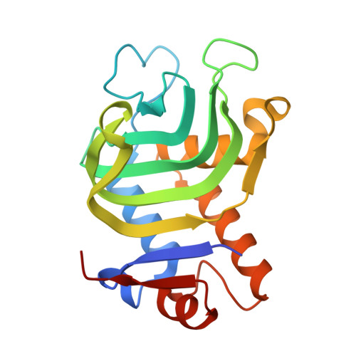
    The heme acquisition system A protein secreted by Pseudomonas aeruginosa (HasA(p)) can capture several synthetic metal complexes other than heme. The crystal structures of HasA(p) harboring synthetic metal complexes revealed only small perturbation of the overall HasA(p) structure. An inhibitory effect upon heme acquisition by HasA(p) bearing synthetic metal complexes was examined by monitoring the growth of Pseudomonas aeruginosa PAO1. HasA(p) bound to iron-phthalocyanine inhibits heme acquisition in the presence of heme-bound HasA(p) as an iron source.


  • Organizational Affiliation

    Department of Chemistry, Graduate School of Science, Nagoya University, Furo-cho, Chikusa-ku, Nagoya 464-8602 (Japan).


Macromolecules
Find similar proteins by:  (by identity cutoff)  |  3D Structure
Entity ID: 1
MoleculeChains Sequence LengthOrganismDetailsImage
Heme acquisition protein HasAp
A, B
186Pseudomonas aeruginosa PAO1Mutation(s): 0 
Gene Names: hasApPA3407
UniProt
Find proteins for G3XD33 (Pseudomonas aeruginosa (strain ATCC 15692 / DSM 22644 / CIP 104116 / JCM 14847 / LMG 12228 / 1C / PRS 101 / PAO1))
Explore G3XD33 
Go to UniProtKB:  G3XD33
Entity Groups  
Sequence Clusters30% Identity50% Identity70% Identity90% Identity95% Identity100% Identity
UniProt GroupG3XD33
Sequence Annotations
Expand
  • Reference Sequence
Experimental Data & Validation

Experimental Data

  • Method: X-RAY DIFFRACTION
  • Resolution: 1.54 Å
  • R-Value Free: 0.194 
  • R-Value Work: 0.175 
  • R-Value Observed: 0.176 
  • Space Group: P 31
Unit Cell:
Length ( Å )Angle ( ˚ )
a = 47.518α = 90
b = 47.518β = 90
c = 140.893γ = 120
Software Package:
Software NamePurpose
DENZOdata reduction
SCALEPACKdata scaling
MOLREPphasing
REFMACrefinement
PDB_EXTRACTdata extraction
HKL-2000data collection

Structure Validation

View Full Validation Report


Ligand Structure Quality Assessment 


Entry History 

Deposition Data

Revision History  (Full details and data files)

  • Version 1.0: 2014-01-29
    Type: Initial release
  • Version 1.1: 2014-04-09
    Changes: Database references
  • Version 1.2: 2023-11-08
    Changes: Data collection, Database references, Derived calculations, Refinement description