3VPL

Crystal structure of a 2-fluoroxylotriosyl complex of the Vibrio sp. AX-4 Beta-1,3-xylanase


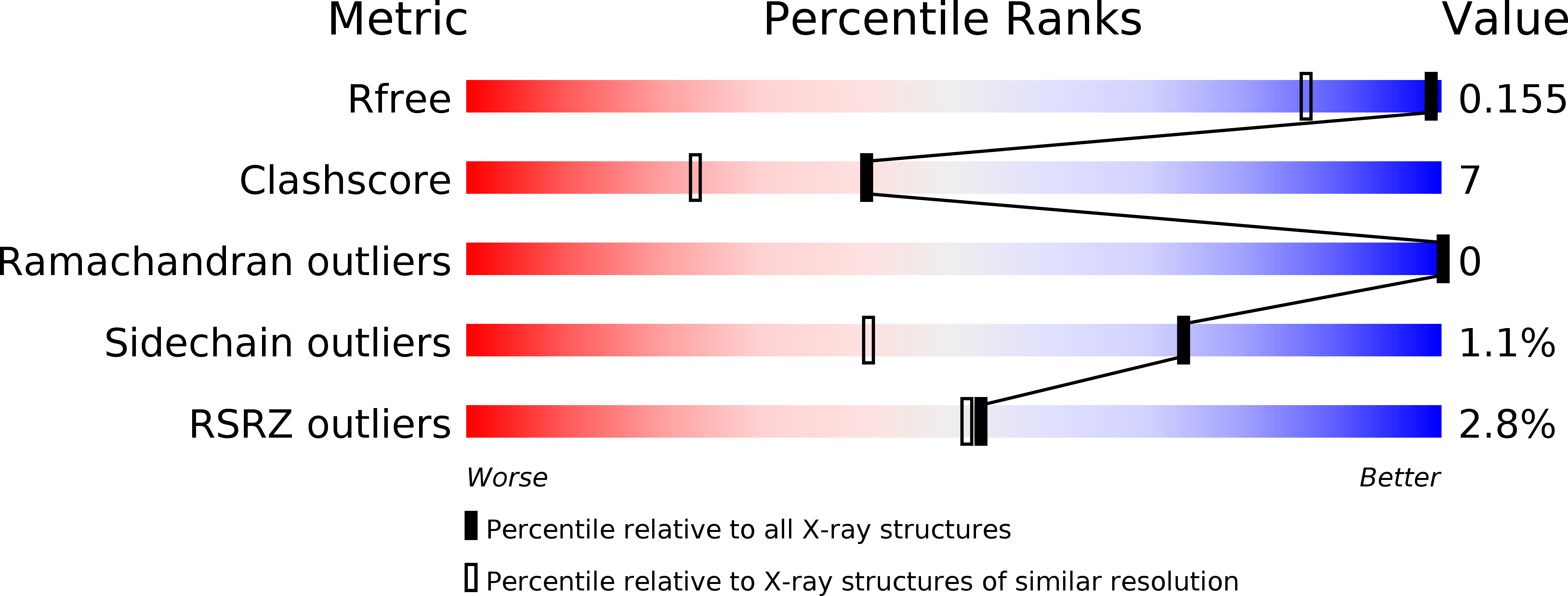
Experimental Data Snapshot

  • Method: X-RAY DIFFRACTION
  • Resolution: 1.20 Å
  • R-Value Free: 0.156 
  • R-Value Work: 0.141 
  • R-Value Observed: 0.142 

wwPDB Validation   3D Report Full Report


Ligand Structure Quality Assessment 


This is version 2.1 of the entry. See complete history


Literature

The crystal structure of a 2-fluoroxylotriosyl complex of the Vibrio sp. AX-4 beta-1,3-xylanase at 1.2 A resolution

Sakaguchi, K.Goddard-Borger, E.D.Kawamura, T.Kiyohara, M.Tanaka, I.Ito, M.Withers, S.G.Watanabe, N.

To be published.

Macromolecules
Find similar proteins by:  (by identity cutoff)  |  3D Structure
Entity ID: 1
MoleculeChains Sequence LengthOrganismDetailsImage
Beta-1,3-xylanase XYL4333Vibrio sp.Mutation(s): 0 
Gene Names: xyl4
EC: 3.2.1.32
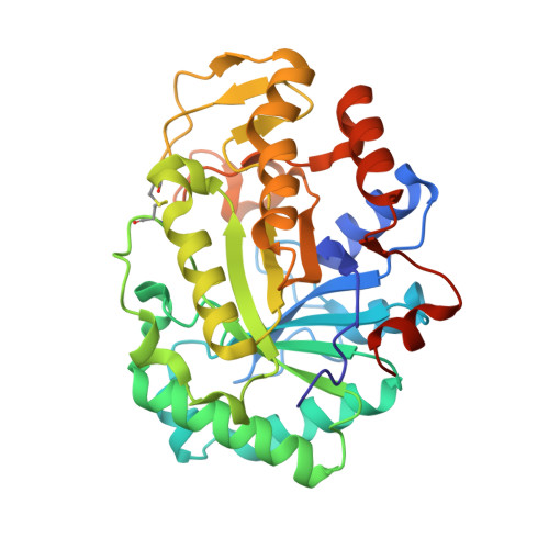
UniProt
Find proteins for D5MP61 (Vibrio sp)
Explore D5MP61 
Go to UniProtKB:  D5MP61
Entity Groups  
Sequence Clusters30% Identity50% Identity70% Identity90% Identity95% Identity100% Identity
UniProt GroupD5MP61
Sequence Annotations
Expand
  • Reference Sequence
Oligosaccharides

Help

Entity ID: 2
MoleculeChains Length2D Diagram Glycosylation3D Interactions
beta-D-xylopyranose-(1-3)-beta-D-xylopyranose-(1-3)-2-deoxy-2-fluoro-beta-D-xylopyranose
B
3N/A
Glycosylation Resources
GlyTouCan:  G18138CS
GlyCosmos:  G18138CS
Entity ID: 3
MoleculeChains Length2D Diagram Glycosylation3D Interactions
beta-D-xylopyranose-(1-4)-beta-D-xylopyranose-(1-3)-2-deoxy-2-fluoro-beta-D-xylopyranose
C
3N/A
Glycosylation Resources
GlyTouCan:  G94440QL
GlyCosmos:  G94440QL
Small Molecules
Ligands 1 Unique
IDChains Name / Formula / InChI Key2D Diagram3D Interactions
DNX
Query on DNX

Download Ideal Coordinates CCD File 
D [auth A]3,4-dinitrophenol
C6 H4 N2 O5
AKLOLDQYWQAREW-UHFFFAOYSA-N
Experimental Data & Validation

Experimental Data

  • Method: X-RAY DIFFRACTION
  • Resolution: 1.20 Å
  • R-Value Free: 0.156 
  • R-Value Work: 0.141 
  • R-Value Observed: 0.142 
  • Space Group: P 21 21 21
Unit Cell:
Length ( Å )Angle ( ˚ )
a = 52.343α = 90
b = 75.437β = 90
c = 81.646γ = 90
Software Package:
Software NamePurpose
SCALEPACKdata scaling
REFMACrefinement
PDB_EXTRACTdata extraction
KEK-PFdata collection
HKL-2000data reduction
MOLREPphasing

Structure Validation

View Full Validation Report


Ligand Structure Quality Assessment 


Entry History 

Deposition Data

Revision History  (Full details and data files)

  • Version 1.0: 2013-03-06
    Type: Initial release
  • Version 2.0: 2020-07-29
    Type: Remediation
    Reason: Carbohydrate remediation
    Changes: Advisory, Atomic model, Data collection, Database references, Derived calculations, Structure summary
  • Version 2.1: 2023-11-08
    Changes: Advisory, Data collection, Database references, Refinement description, Structure summary