3V56

Re-refinement of PDB entry 1OSG - Complex between BAFF and a BR3 derived peptide presented in a beta-hairpin scaffold - reveals an additonal copy of the peptide.


Experimental Data Snapshot

  • Method: X-RAY DIFFRACTION
  • Resolution: 3.00 Å
  • R-Value Free: 0.200 
  • R-Value Work: 0.162 
  • R-Value Observed: 0.165 

wwPDB Validation   3D Report Full Report


This is version 1.3 of the entry. See complete history

Re-refinement Note

This entry reflects an alternative modeling of the original data in: 1OSG


Literature

Exploiting structure similarity in refinement: automated NCS and target-structure restraints in BUSTER.

Smart, O.S.Womack, T.O.Flensburg, C.Keller, P.Paciorek, W.Sharff, A.Vonrhein, C.Bricogne, G.

(2012) Acta Crystallogr D Biol Crystallogr 68: 368-380

  • DOI: https://doi.org/10.1107/S0907444911056058
  • Primary Citation of Related Structures:  
    3SYU, 3URP, 3V56

  • PubMed Abstract: 

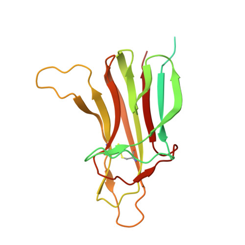
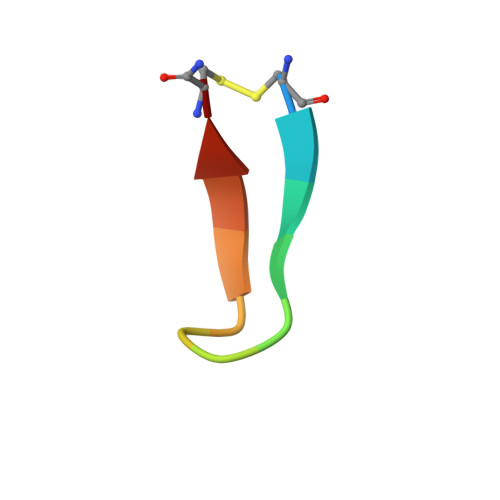
    Maximum-likelihood X-ray macromolecular structure refinement in BUSTER has been extended with restraints facilitating the exploitation of structural similarity. The similarity can be between two or more chains within the structure being refined, thus favouring NCS, or to a distinct 'target' structure that remains fixed during refinement. The local structural similarity restraints (LSSR) approach considers all distances less than 5.5 Å between pairs of atoms in the chain to be restrained. For each, the difference from the distance between the corresponding atoms in the related chain is found. LSSR applies a restraint penalty on each difference. A functional form that reaches a plateau for large differences is used to avoid the restraints distorting parts of the structure that are not similar. Because LSSR are local, there is no need to separate out domains. Some restraint pruning is still necessary, but this has been automated. LSSR have been available to academic users of BUSTER since 2009 with the easy-to-use -autoncs and -target target.pdb options. The use of LSSR is illustrated in the re-refinement of PDB entries 5rnt, where -target enables the correct ligand-binding structure to be found, and 1osg, where -autoncs contributes to the location of an additional copy of the cyclic peptide ligand.


  • Organizational Affiliation

    Global Phasing Ltd, Sheraton House, Castle Park, Cambridge CB3 0AX, England. osmart@globalphasing.com


Macromolecules
Find similar proteins by:  (by identity cutoff)  |  3D Structure
Entity ID: 1
MoleculeChains Sequence LengthOrganismDetailsImage
Tumor necrosis factor ligand superfamily member 13B
A, B, C, D, E
A, B, C, D, E, F
208Homo sapiensMutation(s): 0 
Gene Names: TNFSF13BBAFFBLYSTALL1TNFSF20ZTNF4UNQ401/PRO738
UniProt & NIH Common Fund Data Resources
Find proteins for Q9Y275 (Homo sapiens)
Explore Q9Y275 
Go to UniProtKB:  Q9Y275
PHAROS:  Q9Y275
GTEx:  ENSG00000102524 
Entity Groups  
Sequence Clusters30% Identity50% Identity70% Identity90% Identity95% Identity100% Identity
UniProt GroupQ9Y275
Sequence Annotations
Expand
  • Reference Sequence

Find similar proteins by:  Sequence   |   3D Structure  

Entity ID: 2
MoleculeChains Sequence LengthOrganismDetailsImage
BR3 derived peptive
G,
H,
I,
J,
K,
L,
M [auth Z]
14Homo sapiensMutation(s): 0 
Gene Names: TNFRSF13C
UniProt & NIH Common Fund Data Resources
Find proteins for Q96RJ3 (Homo sapiens)
Explore Q96RJ3 
Go to UniProtKB:  Q96RJ3
PHAROS:  Q96RJ3
GTEx:  ENSG00000159958 
Entity Groups  
Sequence Clusters30% Identity50% Identity70% Identity90% Identity95% Identity100% Identity
UniProt GroupQ96RJ3
Sequence Annotations
Expand
  • Reference Sequence
Experimental Data & Validation

Experimental Data

  • Method: X-RAY DIFFRACTION
  • Resolution: 3.00 Å
  • R-Value Free: 0.200 
  • R-Value Work: 0.162 
  • R-Value Observed: 0.165 
  • Space Group: P 65
Unit Cell:
Length ( Å )Angle ( ˚ )
a = 121.633α = 90
b = 121.633β = 90
c = 157.214γ = 120
Software Package:
Software NamePurpose
AMoREphasing
BUSTERrefinement
DENZOdata reduction
SCALEPACKdata scaling

Structure Validation

View Full Validation Report


Entry History 

Deposition Data

Revision History  (Full details and data files)

  • Version 1.0: 2012-03-28
    Type: Initial release
  • Version 1.1: 2013-02-20
    Changes: Database references
  • Version 1.2: 2018-01-24
    Changes: Database references
  • Version 1.3: 2023-09-13
    Changes: Data collection, Database references, Derived calculations, Refinement description