3U9H

Complex structure of human tankyrase 2 with nicotinamide


Experimental Data Snapshot

  • Method: X-RAY DIFFRACTION
  • Resolution: 1.75 Å
  • R-Value Free: 0.197 
  • R-Value Work: 0.166 
  • R-Value Observed: 0.167 

wwPDB Validation   3D Report Full Report


This is version 1.3 of the entry. See complete history


Literature

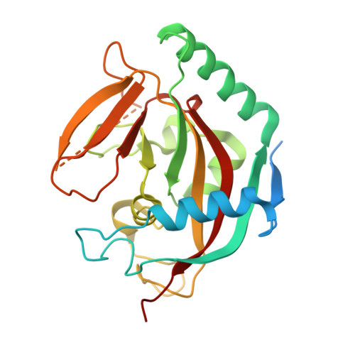
Structural basis of selective inhibition of human tankyrases.

Narwal, M.Venkannagari, H.Lehtio, L.

(2012) J Med Chem 55: 1360-1367

  • DOI: https://doi.org/10.1021/jm201510p
  • Primary Citation of Related Structures:  
    3U9H, 3U9Y, 3UA9

  • PubMed Abstract: 

    Tankyrases are poly(ADP-ribose) polymerases that have many cellular functions. They play pharmaceutically important roles, at least in telomere homeostasis and Wnt signaling, by covalently ADP-ribosylating target proteins and consequently regulating their functions. These features make tankyrases potential targets for treatment of cancer. We report here crystal structures of human tankyrase 2 catalytic fragment in complex with a byproduct, nicotinamide, and with selective inhibitors of tankyrases (IWR-1) and PARPs 1 and 2 (olaparib). Binding of these inhibitors to tankyrase 2 induces specific conformational changes. The crystal structures explain the selectivity of the inhibitors, reveal the flexibility of a substrate binding loop, and explain existing structure-activity relationship data. The first crystal structure of a PARP enzyme in complex with a potent inhibitor, IWR-1, that does not bind to the widely utilized nicotinamide-binding site makes the structure valuable for development of PARP inhibitors in general.


  • Organizational Affiliation

    Pharmaceutical Sciences, Department of Biosciences, Abo Akademi University, FI-20520 Turku, Finland.


Macromolecules
Find similar proteins by:  (by identity cutoff)  |  3D Structure
Entity ID: 1
MoleculeChains Sequence LengthOrganismDetailsImage
Tankyrase-2
A, B
240Homo sapiensMutation(s): 0 
Gene Names: TNKS2PARP5BTANK2TNKL
EC: 2.4.2.30
UniProt & NIH Common Fund Data Resources
Find proteins for Q9H2K2 (Homo sapiens)
Explore Q9H2K2 
Go to UniProtKB:  Q9H2K2
PHAROS:  Q9H2K2
GTEx:  ENSG00000107854 
Entity Groups  
Sequence Clusters30% Identity50% Identity70% Identity90% Identity95% Identity100% Identity
UniProt GroupQ9H2K2
Sequence Annotations
Expand
  • Reference Sequence
Small Molecules
Ligands 5 Unique
IDChains Name / Formula / InChI Key2D Diagram3D Interactions
NCA
Query on NCA

Download Ideal Coordinates CCD File 
F [auth A],
L [auth B]
NICOTINAMIDE
C6 H6 N2 O
DFPAKSUCGFBDDF-UHFFFAOYSA-N
PEG
Query on PEG

Download Ideal Coordinates CCD File 
H [auth A],
M [auth B]
DI(HYDROXYETHYL)ETHER
C4 H10 O3
MTHSVFCYNBDYFN-UHFFFAOYSA-N
SO4
Query on SO4

Download Ideal Coordinates CCD File 
D [auth A],
E [auth A],
J [auth B],
K [auth B]
SULFATE ION
O4 S
QAOWNCQODCNURD-UHFFFAOYSA-L
GOL
Query on GOL

Download Ideal Coordinates CCD File 
G [auth A]GLYCEROL
C3 H8 O3
PEDCQBHIVMGVHV-UHFFFAOYSA-N
ZN
Query on ZN

Download Ideal Coordinates CCD File 
C [auth A],
I [auth B]
ZINC ION
Zn
PTFCDOFLOPIGGS-UHFFFAOYSA-N
Experimental Data & Validation

Experimental Data

  • Method: X-RAY DIFFRACTION
  • Resolution: 1.75 Å
  • R-Value Free: 0.197 
  • R-Value Work: 0.166 
  • R-Value Observed: 0.167 
  • Space Group: C 2 2 21
Unit Cell:
Length ( Å )Angle ( ˚ )
a = 91.25α = 90
b = 97.75β = 90
c = 119.16γ = 90
Software Package:
Software NamePurpose
MxCuBEdata collection
MOLREPphasing
REFMACrefinement
XDSdata reduction
XSCALEdata scaling

Structure Validation

View Full Validation Report


Entry History 

Deposition Data

Revision History  (Full details and data files)

  • Version 1.0: 2011-11-02
    Type: Initial release
  • Version 1.1: 2012-01-25
    Changes: Database references
  • Version 1.2: 2012-02-22
    Changes: Database references
  • Version 1.3: 2023-09-13
    Changes: Data collection, Database references, Derived calculations, Refinement description